You have no items in your shopping cart.
GLI2 Antibody
SKU: orb345484
Description
Images & Validation
−Item 1 of 3
| Tested Applications | ELISA, IHC, WB |
|---|---|
| Dilution range | ELISA: 1:2,000 - 1:12,000, IHC: 1:500 - 1:2,000, WB: 1:500 - 1:2,000 |
| Reactivity | Human, Rat |
| Application Notes |
Key Properties
−| Antibody Type | Primary Antibody |
|---|---|
| Host | Rabbit |
| Clonality | Polyclonal |
| Isotype | IgG |
| Immunogen | This affinity purified antibody was prepared from whole rabbit serum produced by repeated immunizations with a synthetic peptide corresponding to an internal region near amino acids 30-65 of human Gli-2 (isoform a). |
| Purity | This affinity-purified antibody is directed against human Gli-2 protein. The product was affinity purified from monospecific antiserum by immunoaffinity purification. A BLAST analysis was used to suggest cross reactivity with Gli-2 from human and chimpanzee based on the immunizing sequence. |
| Conjugation | Unconjugated |
Storage & Handling
−| Storage | Store vial at -20° C or below prior to opening. This vial contains a relatively low volume of reagent (25 µL). To minimize loss of volume dilute 1:10 by adding 225 µL of the buffer stated above directly to the vial. Recap, mix thoroughly and briefly centrifuge to collect the volume at the bottom of the vial. Use this intermediate dilution when calculating final dilutions as recommended below. Store the vial at -20°C or below after dilution. Avoid cycles of freezing and thawing. |
|---|---|
| Form/Appearance | Liquid (sterile filtered) |
| Buffer/Preservatives | 0.01% (w/v) Sodium Azide |
| Concentration | 1.00 |
| Dry Ice Shipping | Please note: This product requires shipment on dry ice. A dry ice surcharge will apply. |
| Disclaimer | For research use only |
Alternative Names
−rabbit anti-GLI-2 antibody, GLI2, zinc finger protein GLI2, GLI family zinc finger protein 2, Tax helper protein antibody, THP antibody
Similar Products
−- Item 1 of 6
GLI2 Antibody (C-term) [orb1927138]
FC, IF, IHC-P, WB
Human
Rabbit
Polyclonal
Unconjugated
100 μl, 50 μl - Item 1 of 6
Zinc finger protein GLI2 GLI2 Antibody [orb546293]
ELISA, IHC, WB
Human, Mouse, Rat
Rabbit
Polyclonal
Unconjugated
100 μg - Item 1 of 6
- Item 1 of 6
GLI2 Rabbit Polyclonal Antibody [orb329627]
IHC, WB
Human
Human, Mouse
Rabbit
Polyclonal
Unconjugated
100 μl - Item 1 of 6
GLI2 Rabbit Polyclonal Antibody [orb329628]
IHC, WB
Human
Human, Mouse
Rabbit
Polyclonal
Unconjugated
100 μl


Quality Guarantee
Explore bioreagents carefree to elevate your research. All our products are rigorously tested for performance. If a product does not perform as described on its datasheet, our scientific support team will provide expert troubleshooting, a prompt replacement, or a refund. For full details, please see our Terms & Conditions and Buying Guide. Contact us at support@biorbyt.com.
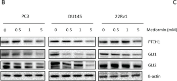
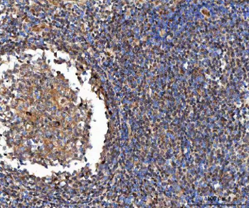
Link between metformin and Hedgehog signaling. (A) GLI1, GLI2 and PTCH1 gene expression after 72-h metformin treatment. Means ± SEM of two independent experiments. * p < 0.05 vs. control; (B) PTCH1, GLI1 and GLI2 protein expression after 72-h metformin treatment; (C) (p)AMPK protein and GLI1 expression in 22Rv1 cells transfected with AMPK siRNA and treated with metformin (5 mM) 72-h prior to protein lysis. GLI1, glioma-associated oncogene homolog 1; GLI2, glioma-associated oncogene homolog 2; PTCH1, patched 1.
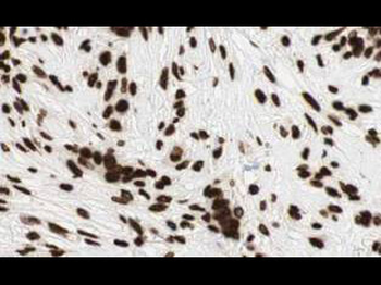
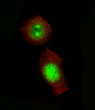
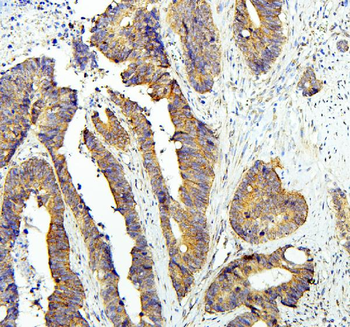
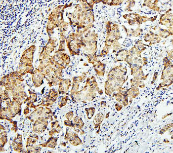
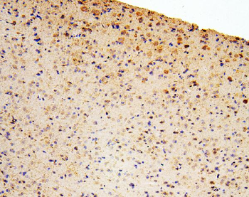
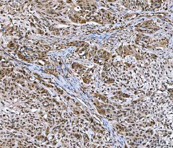
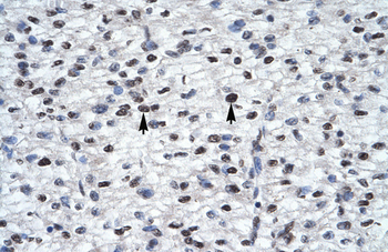
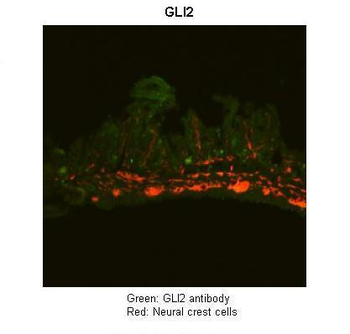
Biorbyt's Affinity Purified anti-Gli2 antibody shows strong cytoplasmic and membranous staining of tumor cells in human breast tissue. Tissue was formalin-fixed and paraffin embedded. Brown color indicates presence of protein, blue color shows cell nuclei.
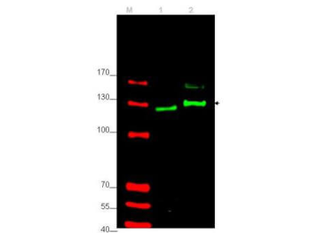
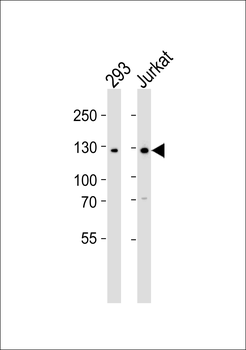
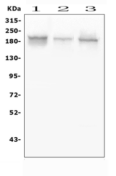
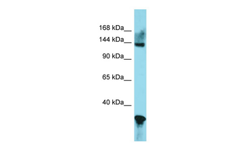
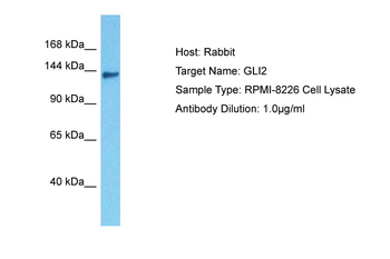
Western blot using Biorbyt's affinity purified anti-Gli-2 antibody shows detection of Gli-2 protein. Lane 1: rat testes and Lane 2: human HEK293 (p/n orb348669) whole cell lysates (arrowhead). Each lane contains approximately 35 µg of lysate. Primary antibody was used at a 1:400 dilution in 5% BLOTTO (p/n orb348624) in PBS overnight at 4°C. The membrane was washed and reacted with a 1:10000 dilution of IRDye® 800 conjugated Gt-a-Rabbit IgG [H&L] MX10 for 45 min at room temperature (800 nm channel, green). Molecular weight estimation was made by comparison to prestained MW markers in lane M (700 nm channel, red).
Quick Database Links
UniProt
RefSeq (Protein):NP_005261.2
UniProt Details
− No UniProt data available
NCBI Reference Sequences
−Associated Accession Numbers
Curated reference sequences for the gene transcript and protein product| Protein | NP_005261.2 |
|---|
Documents Download
Datasheet
Product Information
Request a Document
Protocol Information
WB
Western Blot (IB, immunoblot)
IHC
Immunohistochemistry
ELISA
Enzyme-linked Immunosorbent Assay (EIA)
GLI2 Antibody (orb345484)

- 0.0
Based on 0 reviews
Participating in our Biorbyt product reviews program enables you to support fellow scientists by sharing your firsthand experience with our products.
Login to Submit a Review