You have no items in your shopping cart.
MTHFD1 Antibody
SKU: orb1264604
Featured
Description
Research Area
Cell Biology, Metabolism Research, Signal Transduction
Images & Validation
−Item 1 of 4
| Tested Applications | IF, IHC-P, WB |
|---|---|
| Reactivity | Human |
| Predicted Reactivity | Mouse, Rat |
| Application Notes |
Key Properties
−| Antibody Type | Primary Antibody |
|---|---|
| Host | Rabbit |
| Clonality | Polyclonal |
| Isotype | Rabbit Ig |
| Immunogen | This MTHFD1 antibody is generated from rabbits immunized with a KLH conjugated synthetic peptide between 535-562 amino acids from the Central region of human MTHFD1. |
| Target | MTHFD1 |
| Molecular Weight | 102 kDa |
| Purification | This antibody is prepared by Saturated Ammonium Sulfate (SAS) precipitation followed by dialysis |
| Conjugation | Unconjugated |
Storage & Handling
−| Storage | Maintain refrigerated at 2-8°C for up to 2 weeks. For long term storage store at -20°C in small aliquots to prevent freeze-thaw cycles. |
|---|---|
| Form/Appearance | Liquid |
| Buffer/Preservatives | Supplied in PBS with 0.09% (W/V) sodium azide. |
| Concentration | batch dependent |
| Expiration Date | 12 months from date of receipt. |
| Disclaimer | For research use only |
Alternative Names
−C-1-tetrahydrofolate synthase, cytoplasmic, C1-THF synthase, Methylenetetrahydrofolate dehydrogenase, Methenyltetrahydrofolate cyclohydrolase, Formyltetrahydrofolate synthetase, C-1-tetrahydrofolate synthase, cytoplasmic, N-terminally processed, MTHFD1, MTHFC, MTHFD
Similar Products
−- Item 1 of 10
MTHFD1 Rabbit Polyclonal Antibody [orb1184711]
ELISA, FC, ICC, IF, IHC, WB
Human, Mouse, Rat
Rabbit
Polyclonal
Unconjugated
100 μg - Item 1 of 4
MTHFD1 Antibody (Center P550) [orb1931457]
IF, IHC-P, WB
Mouse, Rat
Human
Rabbit
Polyclonal
Unconjugated
50 μl, 100 μl - Item 1 of 3
- Item 1 of 3
MTHFD1 Antibody [orb629015]
ELISA, IHC, IP, WB
Human, Mouse
Rabbit
Polyclonal
Unconjugated
100 μg, 50 μg - Item 1 of 2
MTHFD1 Rabbit Polyclonal Antibody [orb330471]
IHC, WB
Bovine, Canine, Equine, Guinea pig, Mouse, Rabbit, Rat, Zebrafish
Human
Rabbit
Polyclonal
Unconjugated
100 μl


Quality Guarantee
Explore bioreagents carefree to elevate your research. All our products are rigorously tested for performance. If a product does not perform as described on its datasheet, our scientific support team will provide expert troubleshooting, a prompt replacement, or a refund. For full details, please see our Terms & Conditions and Buying Guide. Contact us at support@biorbyt.com.

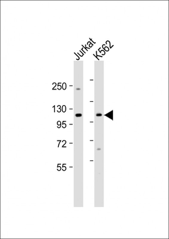
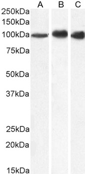
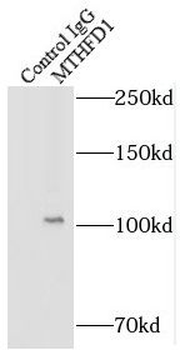
Western Blot at 1:1000 dilution Lane 1: Jurkat whole cell lysate Lane 2: K562 whole cell lysate Lysates/proteins at 20 ug per lane.
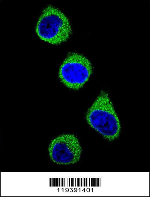
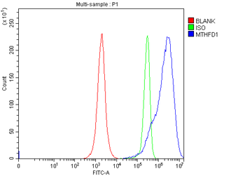
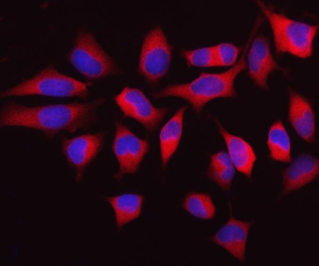
Confocal immunofluorescent analysis of M with 293 cell followed by Alexa Fluor 488-conjugated goat anti-rabbit lgG (green). DAPI was used to stain the cell nuclear (blue).
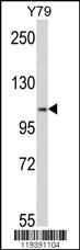
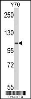
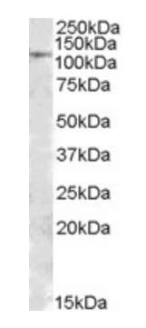
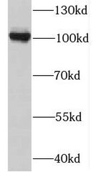
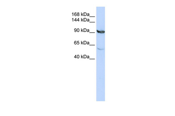
Western blot analysis of M in Y79 cell line lysates (35 ug/lane)
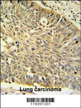
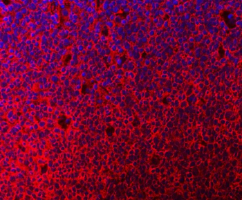
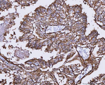
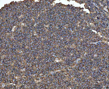
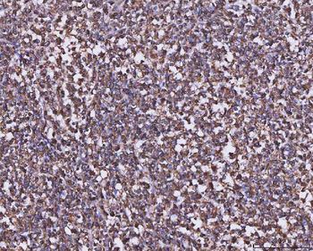
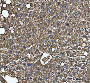
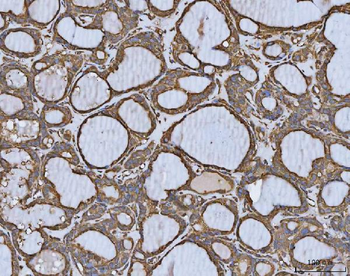
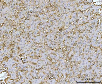
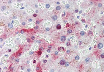
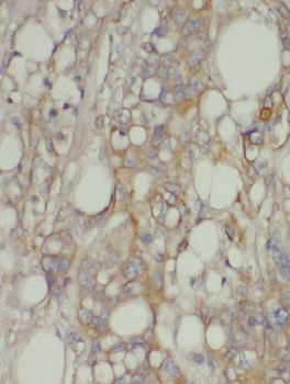
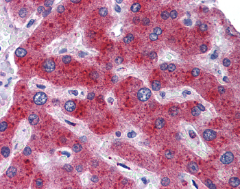
M IHC analysis in formalin fixed and paraffin embedded human Lung carcinoma followed by peroxidase conjugation of the secondary antibody and DAB staining. This data demonstrates the use of the M for immunohistochemistry.
Documents Download
Datasheet
Product Information
Request a Document
Protocol Information
WB
Western Blot (IB, immunoblot)
IHC-P
Immunohistochemistry Paraffin
IF
Immunofluorescence
MTHFD1 Antibody (orb1264604)

- 0.0
Based on 0 reviews
Participating in our Biorbyt product reviews program enables you to support fellow scientists by sharing your firsthand experience with our products.
Login to Submit a ReviewAvailable Sizes
Select a size below