You have no items in your shopping cart.
VDAC1 Antibody (FITC)
SKU: orb150796
Featured
Description
Research Area
Cancer Biology, Neuroscience, Pharmacology & Drug Discovery, Signal Transduction
Images & Validation
−Item 1 of 2
| Tested Applications | ICC, IF, IHC, WB |
|---|---|
| Dilution Range | WB (1:1000) |
| Reactivity | Human, Mouse, Rat |
| Application Notes |
Key Properties
−| Host | Mouse |
|---|---|
| Clonality | Monoclonal |
| Isotype | IgG2a |
| Clone No. | N152B/23 (Formerly sold as S152B-23) |
| Immunogen | Fusion protein amino acids 1-283 (full-length) of human VDAC1. Mouse: 98% identity (279/283 amino acids identical). Rat: 98% identity (279/283 amino acids identical) > 60% identity with VDAC2 and VDAC3. |
| Target | VDAC1 |
| Molecular Weight | 30kDa |
| Purification | Protein G Purified |
| Conjugation | FITC |
Storage & Handling
−| Storage | Conjugated antibodies should be stored according to the product label |
|---|---|
| Buffer/Preservatives | 640.91mM DMSO, 136.36 mM Ethanolamine, 126.89 mM chlorides, 9.09mM phosphates, 9.09mM NaHCO3 |
| Concentration | 1 mg/ml |
| Expiration Date | 12 months from date of receipt. |
| Disclaimer | For research use only |
Alternative Names
−Voltage Dependent Anion Channel 1, Porin, Voltage dependent anion selective channel protein 1, Voltage-dependent anion-selective channel protein 1, hVDAC1, MGC111064, Mitochondrial Porin, Outer mitochondrial membrane protein porin 1, Plasmalemmal porin, Porin 31HL, Porin 31HM, PORIN-31-HL, VDAC 1, VDAC, VDAC-1
Similar Products
−
VDAC1 Rabbit Polyclonal Antibody (FITC) [orb2133913]
IHC, WB
Bovine, Canine, Equine, Guinea pig, Human, Mouse, Rabbit, Rat, Sheep, Zebrafish
Rabbit
Polyclonal
FITC
100 μlVDAC Rabbit Polyclonal Antibody (FITC) [orb16481]
IF
Bovine, Canine, Equine, Mouse, Porcine, Rabbit, Sheep
Human, Rat
Rabbit
Polyclonal
FITC
100 μlVDAC Rabbit Polyclonal Antibody (FITC) [orb526105]
IF
Bovine, Equine, Gallus, Rabbit, Rat, Sheep
Human, Mouse
Rabbit
Polyclonal
FITC
100 μl
Quality Guarantee
Explore bioreagents carefree to elevate your research. All our products are rigorously tested for performance. If a product does not perform as described on its datasheet, our scientific support team will provide expert troubleshooting, a prompt replacement, or a refund. For full details, please see our Terms & Conditions and Buying Guide. Contact us at support@biorbyt.com.

Immunocytochemistry/Immunofluorescence analysis using Mouse Anti-VDAC1 Monoclonal Antibody, Clone N152B/23. Tissue: Neuroblastoma cells (SH-SY5Y). Species: Human. Fixation: 4% PFA for 15 min. Primary Antibody: Mouse Anti-VDAC1 Monoclonal Antibody at 1:100 for overnight at 4°C with slow rocking. Secondary Antibody: AlexaFluor 488 at 1:1000 for 1 hour at RT. Counterstain: Phalloidin-iFluor 647 (red) F-Actin stain; Hoechst (blue) nuclear stain at 1:800, 1.6mM for 20 min at RT. (A) Hoechst (blue) nuclear stain. (B) Phalloidin-iFluor 647 (red) F-Actin stain. (C) VDAC1 Antibody (D) Composite.
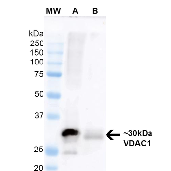
Western Blot analysis of Mouse Brain and Human RT-4 lysates showing detection of ~30 kDa VDAC1 protein using Mouse Anti-VDAC1 Monoclonal Antibody, Clone N152B/23. Lane 1: Molecular Weight Ladder. Lane A: Mouse Brain Lysate. Lane B: Human RT-4 Lysate. Block: 5% Skim milkin TBST. Primary Antibody: Mouse Anti-VDAC1 Monoclonal Antibody at 1:200 for 60 min at RT. Secondary Antibody: Goat Anti-Mouse IgG: HRP at 1:2000 for 60 min at RT. Color Development: ECL solution for 5 min in RT. Predicted/Observed Size: ~30 kDa.
Quick Database Links
UniProt Details
− No UniProt data available
NCBI Gene Details
− No NCBI Gene data available
NCBI Reference Sequences
−Associated Accession Numbers
Curated reference sequences for the gene transcript and protein product| Protein | NP_003365.1 |
|---|
Documents Download
Datasheet
Product Information
Request a Document
Protocol Information
WB
Western Blot (IB, immunoblot)
IHC
Immunohistochemistry
IF
Immunofluorescence
ICC
Immunocytochemistry
VDAC1 Antibody (FITC) (orb150796)

- 0.0
Based on 0 reviews
Participating in our Biorbyt product reviews program enables you to support fellow scientists by sharing your firsthand experience with our products.
Login to Submit a ReviewAvailable Sizes
Select a size below