You have no items in your shopping cart.
ABCG2 Recombinant Rabbit Monoclonal Antibody
SKU: orb1499376
Featured
Description
Research Area
ATPases, Cancer Research, Drug Metabolism, Metabolism, Molecular Biology, Signal Transduction, Surface Molecules, Tumor Biology
Images & Validation
−Item 1 of 4
| Tested Applications | IF, IHC-Fr, IHC-P, WB |
|---|---|
| Dilution Range | WB=1:500-2000, IHC-P=1:100-500, IHC-F=1:100-500, IF=1:100-500 |
| Reactivity | Human |
| Predicted Reactivity | Human, Mouse |
Key Properties
−| Antibody Type | Primary Antibody |
|---|---|
| Host | Rabbit |
| Clonality | Recombinant |
| Isotype | IgG |
| Clone No. | 4C4 |
| Immunogen | A synthesized peptide derived from human ABCG2 (150-200/655aa) |
| Target | ABCG2 |
| Molecular Weight | 72 kDa |
| Purification | Affinity purified by Protein A |
| Conjugation | Unconjugated |
Storage & Handling
−| Storage | Maintain refrigerated at 2-8°C for up to 2 weeks. For long term storage store at -20°C in small aliquots to prevent freeze-thaw cycles. |
|---|---|
| Form/Appearance | Liquid |
| Buffer/Preservatives | 0.01M TBS (pH7.4) with 1% rAlbumin, 0.02% Proclin300 and 50% Glycerol. |
| Concentration | 1mg/ml |
| Expiration Date | 12 months from date of receipt. |
| Disclaimer | For research use only |
Alternative Names
−ABC15; ABCG 2; ABCG2_HUMAN; ABCP; ATP binding cassette sub family G(WHITE) member 2; ATP binding cassette transporter G2; ATP-binding cassette sub-family G member 2; BCRP1; BMDP; Breast cancer resistance protein; CD338; CDw338; CDw338 antigen; EST157481; GOUT1; MGC102821; Mitoxantrone resistance associated protein; Mitoxantrone resistance-associated protein; MRX; Multi drug resistance efflux transport ATP binding cassette sub family G(WHITE) member 2; MXR; MXR1; Placenta specific ATP binding cassette transporter; Placenta specific MDR protein; Placenta-specific ATP-binding cassette transporter; UAQTL1.
Quality Guarantee
Explore bioreagents carefree to elevate your research. All our products are rigorously tested for performance. If a product does not perform as described on its datasheet, our scientific support team will provide expert troubleshooting, a prompt replacement, or a refund. For full details, please see our Terms & Conditions and Buying Guide. Contact us at support@biorbyt.com.
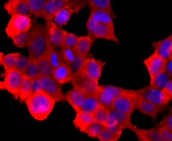
ICC staining of ABCG2 in 293T cells (red). Formalin fixed cells were permeabilized with 0.1% Triton X-100 in TBS for 10 minutes at room temperature and blocked with 1% Blocker BSA for 15 minutes at room temperature. Cells were probed with the primary antibody (orb1499376, 1/50) for 1 hour at room temperature, washed with PBS. Alexa Fluor®594 Goat anti-Rabbit IgG was used as the secondary antibody at 1/1000 dilution. The nuclear counter stain is DAPI (blue).
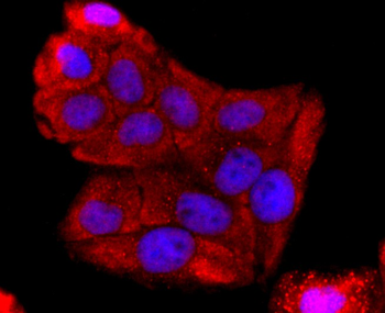
ICC staining of ABCG2 in Hela cells (red). Formalin fixed cells were permeabilized with 0.1% Triton X-100 in TBS for 10 minutes at room temperature and blocked with 1% Blocker BSA for 15 minutes at room temperature. Cells were probed with the primary antibody (orb1499376, 1/50) for 1 hour at room temperature, washed with PBS. Alexa Fluor®594 Goat anti-Rabbit IgG was used as the secondary antibody at 1/1000 dilution. The nuclear counter stain is DAPI (blue).
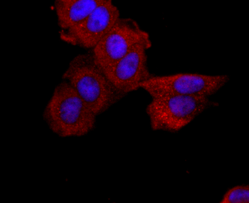
ICC staining of ABCG2 in MCF-7 cells (red). Formalin fixed cells were permeabilized with 0.1% Triton X-100 in TBS for 10 minutes at room temperature and blocked with 1% Blocker BSA for 15 minutes at room temperature. Cells were probed with the primary antibody (orb1499376, 1/50) for 1 hour at room temperature, washed with PBS. Alexa Fluor®594 Goat anti-Rabbit IgG was used as the secondary antibody at 1/1000 dilution. The nuclear counter stain is DAPI (blue).
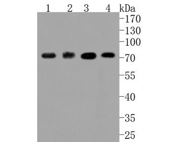
Western blot analysis of ABCG2 on different lysates. Proteins were transferred to a PVDF membrane and blocked with 5% BSA in PBS for 1 hour at room temperature. The primary antibody (orb1499376, 1/500) was used in 5% BSA at room temperature for 2 hours. Goat Anti-Rabbit IgG - HRP Secondary Antibody (HA1001) at 1:5000 dilution was used for 1 hour at room temperature. Positive control: Lane 1: HepG2 cell lysate, Lane 2: human placenta tissue lysate, Lane 3: Hela cell lysate, Lane 4: 293T cell lysate.
Quick Database Links
Gene Symbol
ABCG2
UniProt
UniProt Details
− No UniProt data available
Documents Download
Datasheet
Product Information
Request a Document
Protocol Information
WB
Western Blot (IB, immunoblot)
IHC-P
Immunohistochemistry Paraffin
IHC-Fr
Immunohistochemistry Frozen
IF
Immunofluorescence
ABCG2 Recombinant Rabbit Monoclonal Antibody (orb1499376)

- 0.0
Based on 0 reviews
Participating in our Biorbyt product reviews program enables you to support fellow scientists by sharing your firsthand experience with our products.
Login to Submit a ReviewAvailable Sizes
Select a size below