You have no items in your shopping cart.
Description
Research Area
Infectious Disease & Virology, Stem Cell & Developmental Biology
Images & Validation
−Item 1 of 3
| Tested Applications | WB |
|---|---|
| Dilution Range | WB: 1:1000, WB: 1:1000, WB: 1:1000 |
| Reactivity | Human |
| Predicted Reactivity | Mouse, Zebrafish |
Key Properties
−| Antibody Type | Primary Antibody |
|---|---|
| Host | Rabbit |
| Clonality | Polyclonal |
| Isotype | Rabbit IgG |
| Immunogen | This AGR2 antibody is generated from rabbits immunized with a KLH conjugated synthetic peptide between 95-124 amino acids from the Central region of human AGR2. Antigen Region: 95-124 aa. |
| Target | AGR2 |
| Molecular Weight | 19979 Da |
| Conjugation | Unconjugated |
Storage & Handling
−| Storage | Maintain refrigerated at 2-8°C for up to 2 weeks. For long term storage store at -20°C in small aliquots to prevent freeze-thaw cycles |
|---|---|
| Form/Appearance | Purified polyclonal antibody supplied in PBS with 0.09% (W/V) sodium azide. This antibody is purified through a protein A column, followed by peptide affinity purification. |
| Expiration Date | 12 months from date of receipt. |
| Disclaimer | For research use only |
Alternative Names
−AGR2; AG2; Anterior gradient protein 2 homolog; HPC8; Secreted cement gland protein XAG-2 homolog
Similar Products
−- Item 1 of 3
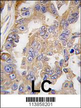
AGR2 Antibody (Center) [orb1930126]
IHC-P, WB
Zebrafish
Human, Mouse
Rabbit
Polyclonal
Unconjugated
100 μl, 50 μl

Quality Guarantee
Explore bioreagents carefree to elevate your research. All our products are rigorously tested for performance. If a product does not perform as described on its datasheet, our scientific support team will provide expert troubleshooting, a prompt replacement, or a refund. For full details, please see our Terms & Conditions and Buying Guide. Contact us at support@biorbyt.com.
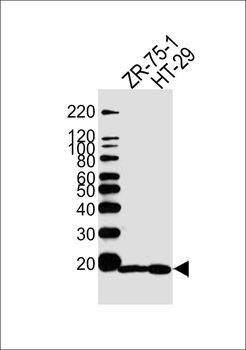
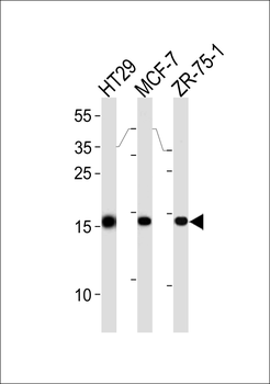
Western blot analysis of lysates from ZR-75-1, HT29 cell line (from left to right), using AGR2 Antibody (Center). Diluted at 1:1000 at each lane. A goat anti-rabbit IgG H&L (HRP) at 1:5000 dilution was used as the secondary antibody.
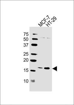
Western blot analysis of lysates from MCF-7, HT-29 cell line (from left to right), using AGR2 Antibody (Center). Diluted at 1:1000 at each lane. A goat anti-rabbit IgG H&L (HRP) at 1:10000 dilution was used as the secondary antibody.
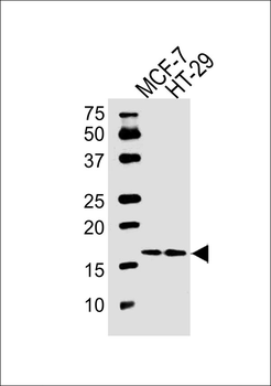
Western blot analysis of lysates from MCF-7, HT-29 cell line (from left to right), using AGR2 Antibody (Center). Diluted at 1:1000 at each lane. A goat anti-rabbit IgG H&L (HRP) at 1:10000 dilution was used as the secondary antibody.
Quick Database Links
UniProt Details
− No UniProt data available
NCBI Reference Sequences
−Associated Accession Numbers
Curated reference sequences for the gene transcript and protein product| Protein | NP_006399.1 |
|---|
Documents Download
Datasheet
Product Information
Request a Document
AGR2 Antibody (Center) (orb1788328)

- 0.0
Based on 0 reviews
Participating in our Biorbyt product reviews program enables you to support fellow scientists by sharing your firsthand experience with our products.
Login to Submit a Review