You have no items in your shopping cart.
AKT1 Antibody
SKU: orb344406
Description
Images & Validation
−Item 1 of 2
| Tested Applications | ELISA, IF, IHC, WB |
|---|---|
| Dilution Range | ELISA: 1:20,000, IHC: 20 µg/ml, IF: 1:25, WB: 1:500 - 1:3,000 |
| Reactivity | Human, Mouse |
| Application Notes |
Key Properties
−| Antibody Type | Primary Antibody |
|---|---|
| Host | Mouse |
| Clonality | Monoclonal |
| Isotype | IgG1 |
| Clone No. | 18F3.H11 |
| Immunogen | Anti-AKT pT308 monoclonal antibody was produced by repeated immunizations with a synthetic peptide corresponding to residues surrounding T308 of human AKT1 protein. |
| Target | AKT1 |
| Purity | Anti-AKT Phospho pT308 antibody was purified from concentrated tissue culture supernate by Protein A chromatography. This antibody is specific for human and mouse AKT protein phosphorylated at T308. A BLAST analysis was used to suggest cross-reactivity with AKT pT308 from most vertebrate species sources based on 100% homology with the immunizing sequence. Cross-reactivity with AKT from other sources has not been determined. Cross-reactivity with AKT2 and AKT3 will likely occur. Cancer, Cell Signaling, Neuroscience, Signal Transduction research. |
| Conjugation | Unconjugated |
Storage & Handling
−| Storage | Store vial at -20° C or below prior to opening. This vial contains a relatively low volume of reagent (25 µL). To minimize loss of volume dilute 1:10 by adding 225 µL of the buffer stated above directly to the vial. Recap, mix thoroughly and briefly centrifuge to collect the volume at the bottom of the vial. Use this intermediate dilution when calculating final dilutions as recommended below. Store the vial at -20°C or below after dilution. Avoid cycles of freezing and thawing. |
|---|---|
| Form/Appearance | Liquid (sterile filtered) |
| Buffer/Preservatives | Preservative: 0.01% (w/v) Sodium Azide. Stabilizer: None; Buffer: 0.02 M Potassium Phosphate, 0.15 M Sodium Chloride, pH 7.2 |
| Concentration | 1.0 mg/ml |
| Expiration Date | 12 months from date of receipt. |
| Dry Ice Shipping | Please note: This product requires shipment on dry ice. A dry ice surcharge will apply. |
| Disclaimer | For research use only |
Alternative Names
−mouse anti-AKT pT308 Antibody, RAC-PK-alpha, Protein kinase B, PKB, C-AKT, RAC-alpha serine/threonine-protein kinase, Proto-oncogene c-Akt, AKT1, AKT 1, AKT-1
Similar Products
−- Item 1 of 11
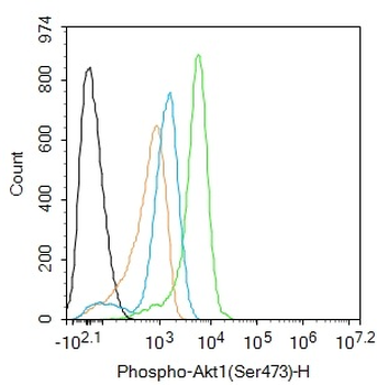
Phospho-AKT (Ser473) Rabbit Polyclonal Antibody [orb11293]
WB
Mouse, Rat
Human
Rabbit
Polyclonal
Unconjugated
50 μl, 100 μl, 200 μl - Item 1 of 9
AKT1 Antibody [orb344403]
ELISA, IF, IHC, Multiplex Assay, WB
Human, Monkey, Mouse, Rat
Mouse
Monoclonal
Unconjugated
100 μg - Item 1 of 9
AKT1 Mouse Polyclonal Antibody [orb11276]
ICC, IF, IHC-Fr, IHC-P, WB
Bovine, Canine, Gallus, Porcine, Rabbit, Sheep
Human, Mouse, Rat
Mouse
Polyclonal
Unconjugated
50 μl, 100 μl, 200 μl - Item 1 of 11
Phospho-Akt (Thr450) Recombinant Rabbit Monoclonal Antibody [orb559195]
IF, IHC-Fr, IHC-P
Mouse, Rat
Human, Mouse, Rat
Rabbit
Recombinant
Unconjugated
25 μl, 100 μl, 50 μl - Item 1 of 8
Phospho-Akt1 (Ser473) Recombinant Rabbit Monoclonal Antibody [orb526646]
WB
Mouse, Rat
Human
Rabbit
Recombinant
Unconjugated
50 μl, 100 μl, 25 μl


Quality Guarantee
Explore bioreagents carefree to elevate your research. All our products are rigorously tested for performance. If a product does not perform as described on its datasheet, our scientific support team will provide expert troubleshooting, a prompt replacement, or a refund. For full details, please see our Terms & Conditions and Buying Guide. Contact us at support@biorbyt.com.
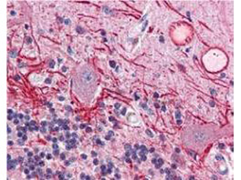
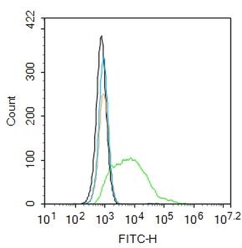
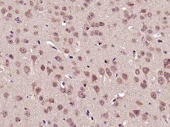
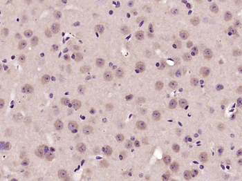
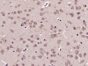
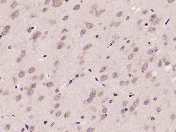
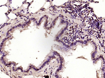
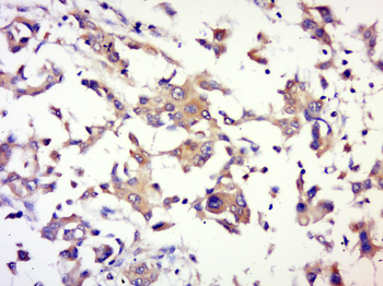
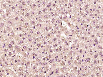
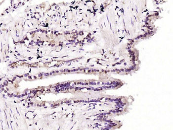
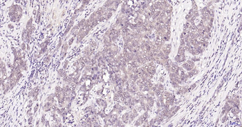
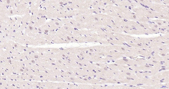
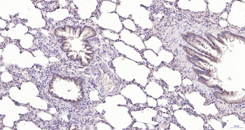
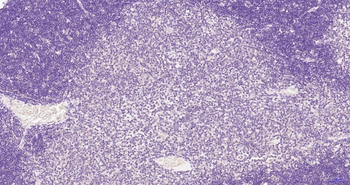
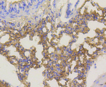
Immunohistochemistry of Mouse anti-AKT pT308 antibody. Tissue: human brain cerebellum tissue (40X). Fixation: formalin fixed paraffin embedded. Antigen retrieval: not required. Primary antibody: AKT pT308antibody at 20 µg/ml for 1 h at RT. Secondary antibody: Peroxidase rabbit secondary antibody at 1:10000 for 45 min at RT. Localization: staining of Purkinje neurons and cell processes in the cerebellum, cytosolic as well as occasionally nuclear. Staining: AKT pT308 as precipitated red signal with hematoxylin purple nuclear counterstain.
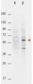
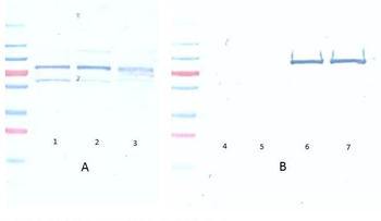
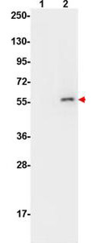
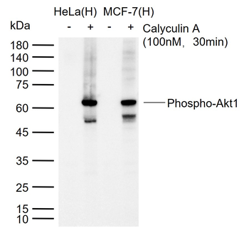
Western Blot of Mouse anti-AKT pT308 antibody. Lane 1: non-phosphorylated AKT in untreated NIH/3T3 cells (p/n orb348714). Lane 2: phosphorylated AKT on PDGF stimulated NIH/3T3 cell lysates (p/n orb348723). Load: 15 µg per lane. Primary antibody: AKT pT308 antibody at a 1:4000 dilution in TBS with 3% BSA, for 3 h at 4°C. Secondary antibody: Peroxidase conjugated Gt-a-Mouse IgG (Fc) (p/n orb347428) was used at a 1:40000 dilution for 1 h at 4°C. Block: 3% BSA in TBS for 30 min at RT. Predicted/Observed size: (indicated by arrowhead at ~56 kDa). Other band(s): unspecific.
Documents Download
Datasheet
Product Information
Request a Document
Protocol Information
WB
Western Blot (IB, immunoblot)
IHC
Immunohistochemistry
IF
Immunofluorescence
ELISA
Enzyme-linked Immunosorbent Assay (EIA)
AKT1 Antibody (orb344406)

- 0.0
Based on 0 reviews
Participating in our Biorbyt product reviews program enables you to support fellow scientists by sharing your firsthand experience with our products.
Login to Submit a ReviewAvailable Sizes
Select a size below