You have no items in your shopping cart.
Description
Images & Validation
−Item 1 of 4
| Tested Applications | IHC-P, IP, WB |
|---|---|
| Reactivity | Human |
| Application Notes |
Key Properties
−| Antibody Type | Primary Antibody |
|---|---|
| Clonality | Monoclonal |
| Isotype | Mouse IgG1 |
| Clone No. | T2H5 |
| Immunogen | Protein preparation from a homogenate of a human mammary tumour specimen. |
| Target | Tenascin C |
| Purification | Purified by protein-A affinity chromatography. |
| Conjugation | Unconjugated |
Storage & Handling
−| Storage | Maintain refrigerated at 2-8°C for up to 2 weeks. For long term storage store at -20°C in small aliquots to prevent freeze-thaw cycles. |
|---|---|
| Buffer/Preservatives | Tris buffered saline (TBS), pH 8.0, 15 mM sodium azide |
| Concentration | 1 mg/ml |
| Expiration Date | 12 months from date of receipt. |
| Disclaimer | For research use only |
Alternative Names
−TNC, Hexabrachion, Cytotactin, Neuronectin
Similar Products
−- Item 1 of 4
Tenascin-C rabbit pAb Antibody [orb767153]
ELISA, IF, IHC, WB
Human, Mouse, Rat
Polyclonal
Unconjugated
50 μl, 100 μl - Item 1 of 1
- Item 1 of 2
Tenascin C Rabbit Polyclonal Antibody [orb11493]
IF, IHC-Fr, IHC-P
Bovine, Canine, Gallus, Human, Porcine, Rabbit
Mouse, Rat
Rabbit
Polyclonal
Unconjugated
50 μl, 100 μl, 200 μl - Item 1 of 2
Tenascin C Rabbit Polyclonal Antibody [orb11461]
IF, IHC-Fr, IHC-P
Human, Porcine
Mouse, Rat
Rabbit
Polyclonal
Unconjugated
50 μl, 100 μl, 200 μl 


Quality Guarantee
Explore bioreagents carefree to elevate your research. All our products are rigorously tested for performance. If a product does not perform as described on its datasheet, our scientific support team will provide expert troubleshooting, a prompt replacement, or a refund. For full details, please see our Terms & Conditions and Buying Guide. Contact us at support@biorbyt.com.
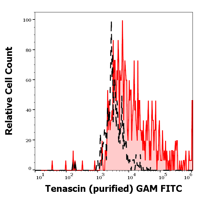
Separation of U-87 MG cells stained using anti-tenascin C (T2H5) purified antibody (concentration in sample 12 µg/ml, GAM FITC, red-filled) from U-87 MG cells unstained by primary antibody (GAM FITC, black-dashed) in flow cytometry analysis (surface staining).
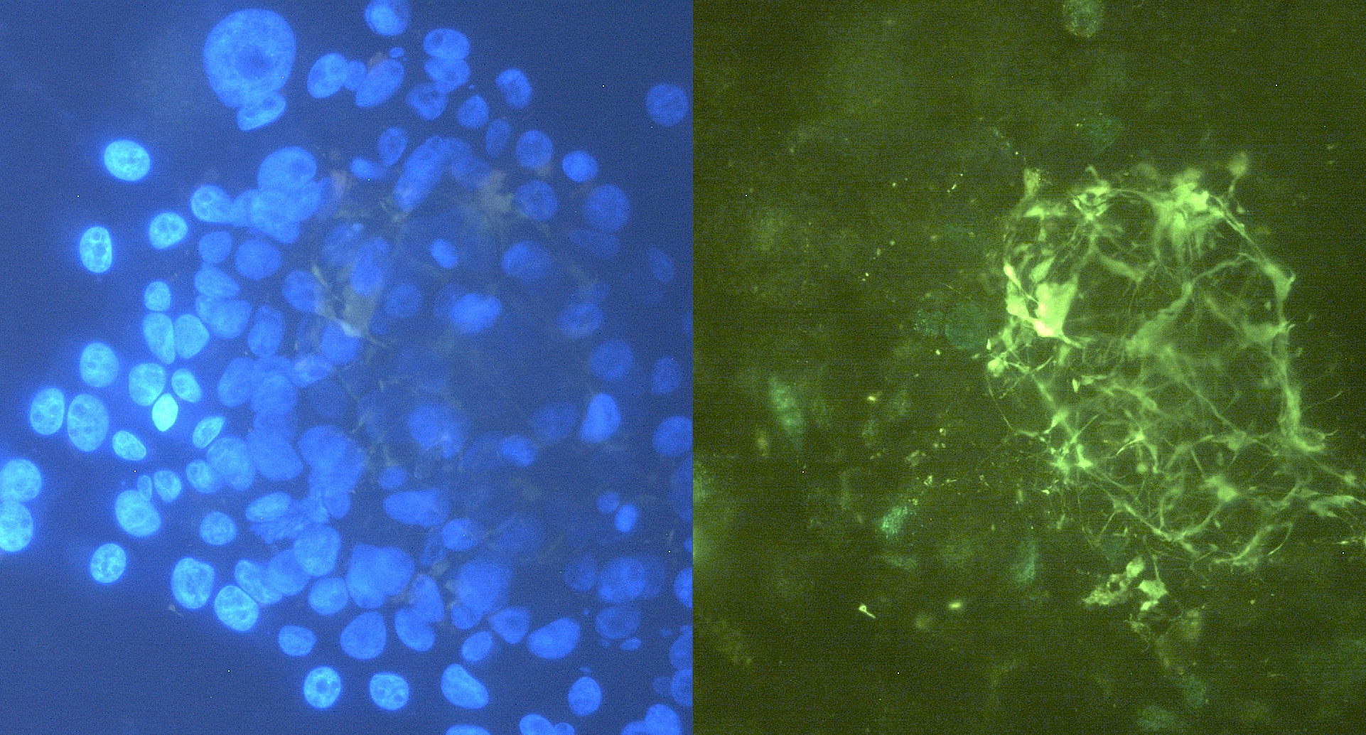
Immunocytochemistry staining of tenascin C in U-87 MG cells using purified mouse monoclonal antibody T2H7 (concentration in sample 12 µg/ml, GAM FITC, right picture) vs. Hoechst 34580 nuclear staining (left picture).

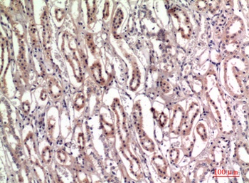
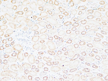
Immunohistochemistry staining of tonsil (paraffin-embedded sections) with anti-human tenascin C (T2H5).
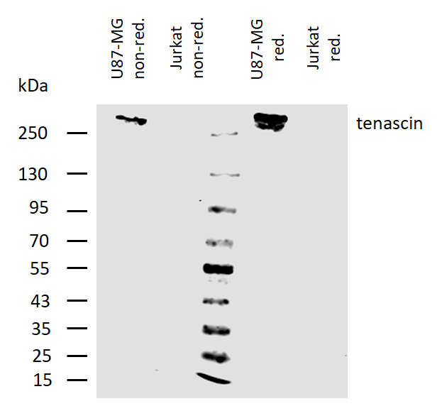
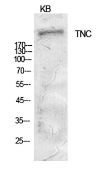
Western blotting analysis of human tenascin C using mouse monoclonal antibody T2H5 on lysates of U87-MG cell line and Jurkat cell line (tenascin non-expressing cell line; negative control) under non-reducing and reducing conditions. Cells were lysed by 50 mM TRIS-Cl pH 6.6, 4M urea, 4% SDS, samples were mixed and heated (100°C, 5 min) with reducing and non-reducing SDS-loading buffer, then resolved using 7.5% Tris-glycine SDS gel electrophoresis. Nitrocellulose membrane was probed with 2 µg/ml of mouse anti-tenascin monoclonal antibody followed by IRDye800-conjugated anti-mouse secondary antibody. Tenascin C was detected slightly above 250 kDa.
Documents Download
Datasheet
Product Information
Request a Document
Protocol Information
WB
Western Blot (IB, immunoblot)
IHC-P
Immunohistochemistry Paraffin
IP
Immunoprecipitation
Tenascin C Antibody (orb44503)

- 0.0
Based on 0 reviews
Participating in our Biorbyt product reviews program enables you to support fellow scientists by sharing your firsthand experience with our products.
Login to Submit a ReviewAvailable Sizes
Select a size below
Free Secondary Antibody (20 ul)0/0
Please add an antibody product to your cart first.