You have no items in your shopping cart.
LCK (Phospho-Y393) Rabbit Polyclonal Antibody
SKU: orb214180
Featured
Description
Research Area
Cancer, Epigenetics, Infectious Diseases
Images & Validation
−Item 1 of 3
| Tested Applications | WB |
|---|---|
| Dilution Range | WB: 1-500-1-1000, IF/ICC: 1-100-1-500 |
| Reactivity | Bovine, Gallus, Human, Mouse, Porcine, Rat, Sheep |
Key Properties
−| Antibody Type | Primary Antibody |
|---|---|
| Host | Rabbit |
| Clonality | Polyclonal |
| Immunogen | KLH-conjugated synthetic phosphopeptide corresponding to residues surrounding Y393 of human LCK protein. The exact sequence is proprietary. |
| Target | LCK |
| Purification | The antibody was purified by immunogen affinity chromatography. |
| Conjugation | Unconjugated |
Storage & Handling
−| Storage | Maintain refrigerated at 2-8°C for up to 2 weeks. For long term storage store at -20°C in small aliquots to prevent freeze-thaw cycles. |
|---|---|
| Form/Appearance | Liquid |
| Buffer/Preservatives | 0.42% Potassium phosphate, 0.87% Sodium chloride, pH 7.3, 30% glycerol, and 0.01% sodium azide. |
| Expiration Date | 12 months from date of receipt. |
| Disclaimer | For research use only |
Alternative Names
−Tyrosine-protein kinase Lck; Leukocyte C-terminal Src kinase; LSK; Lymphocyte cell-specific protein-tyrosine kinase; Protein YT16; Proto-oncogene Lck; T cell-specific protein-tyrosine kinase; p56-LCK
Quality Guarantee
Explore bioreagents carefree to elevate your research. All our products are rigorously tested for performance. If a product does not perform as described on its datasheet, our scientific support team will provide expert troubleshooting, a prompt replacement, or a refund. For full details, please see our Terms & Conditions and Buying Guide. Contact us at support@biorbyt.com.
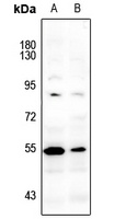
Western blot analysis of LCK (Phospho-Y393) expression in Myla2059 (A), HuT78 (B) whole cell lysates. (Predicted band size: 58 kD; Observed band size: 56 kD)
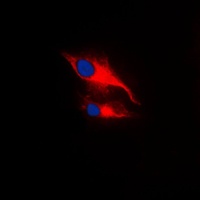
Immunofluorescent analysis of LCK (Phospho-Y393) staining in MCF7 cells. Formalin-fixed cells were permeabilized with 0.1% Triton X-100 in TBS for 5-10 minutes and blocked with 3% BSA-PBS for 30 minutes at room temperature. Cells were probed with the primary antibody in 3% BSA-PBS and incubated overnight at 4 °C in a humidified chamber. Cells were washed with PBST and incubated with a DyLight 594-conjugated secondary antibody (red) in PBS at room temperature in the dark. DAPI was used to stain the cell nuclei (blue).
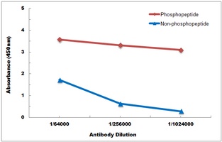
Direct ELISA antibody dose-response curve using Anti-LCK (Phospho-Y393) Antibody. Antigen (Phosphopeptide and non-phosphopeptide) concentration is 5 ug/ml. Goat Anti-Rabbit IgG (H&L) - HRP was used as the secondary antibody, and signal was developed by TMB substrate.
Documents Download
Datasheet
Product Information
Request a Document
LCK (Phospho-Y393) Rabbit Polyclonal Antibody (orb214180)

- 0.0
Based on 0 reviews
Participating in our Biorbyt product reviews program enables you to support fellow scientists by sharing your firsthand experience with our products.
Login to Submit a ReviewAvailable Sizes
Select a size below