You have no items in your shopping cart.
SIT Antibody
SKU: orb44488
Description
Research Area
Epigenetics, Immunology
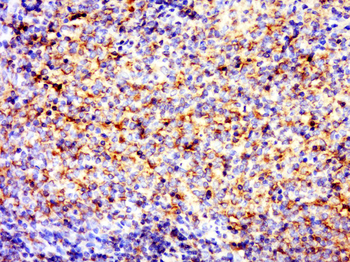
Images & Validation
−Item 1 of 6
| Tested Applications | FC, IP, WB |
|---|---|
| Reactivity | Human |
| Application Notes |
Key Properties
−| Antibody Type | Primary Antibody |
|---|---|
| Clonality | Monoclonal |
| Isotype | Mouse IgG1 |
| Clone No. | SIT-01 |
| Immunogen | Bacterially produced recombinant intracellular fragment of human SIT. |
| Target | SIT |
| Purification | Purified by protein-A affinity chromatography. |
| Conjugation | Unconjugated |
Storage & Handling
−| Storage | Maintain refrigerated at 2-8°C for up to 2 weeks. For long term storage store at -20°C in small aliquots to prevent freeze-thaw cycles. |
|---|---|
| Buffer/Preservatives | Phosphate buffered saline (PBS), pH 7.4, 15 mM sodium azide |
| Concentration | 1 mg/ml |
| Expiration Date | 12 months from date of receipt. |
| Disclaimer | For research use only |
Alternative Names
−SIT1
Similar Products
−- Item 1 of 1
- Item 1 of 1
- Item 1 of 1
SIT Rabbit Polyclonal Antibody [orb158392]
IF, IHC-Fr, IHC-P
Bovine, Human, Mouse, Porcine, Sheep
Rat
Rabbit
Polyclonal
Unconjugated
50 μl, 100 μl, 200 μl - Item 1 of 1
Quality Guarantee
Explore bioreagents carefree to elevate your research. All our products are rigorously tested for performance. If a product does not perform as described on its datasheet, our scientific support team will provide expert troubleshooting, a prompt replacement, or a refund. For full details, please see our Terms & Conditions and Buying Guide. Contact us at support@biorbyt.com.
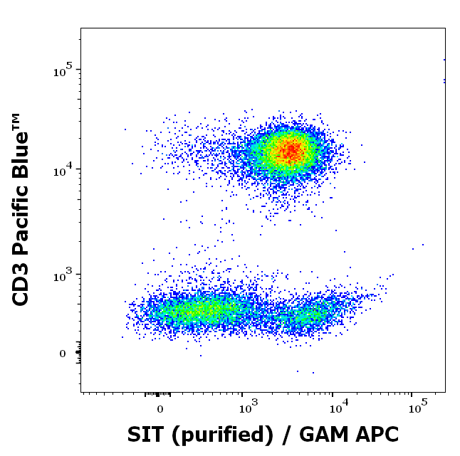
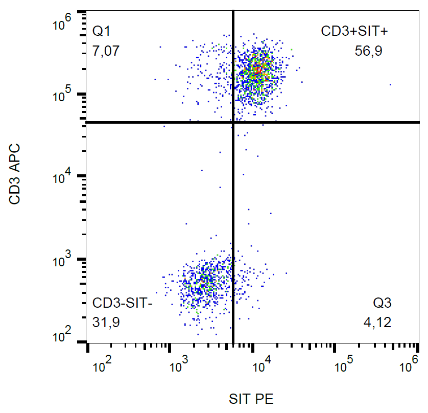
Flow cytometry multicolor intracellular staining of human peripheral whole blood stained using anti-SIT (SIT-01) purified antibody (concentration in sample 9 µg/ml, GAM APC) and anti-human CD3 (UCHT1) Pacific Blue™ antibody (20 µl reagent / 100 µl of peripheral whole blood).
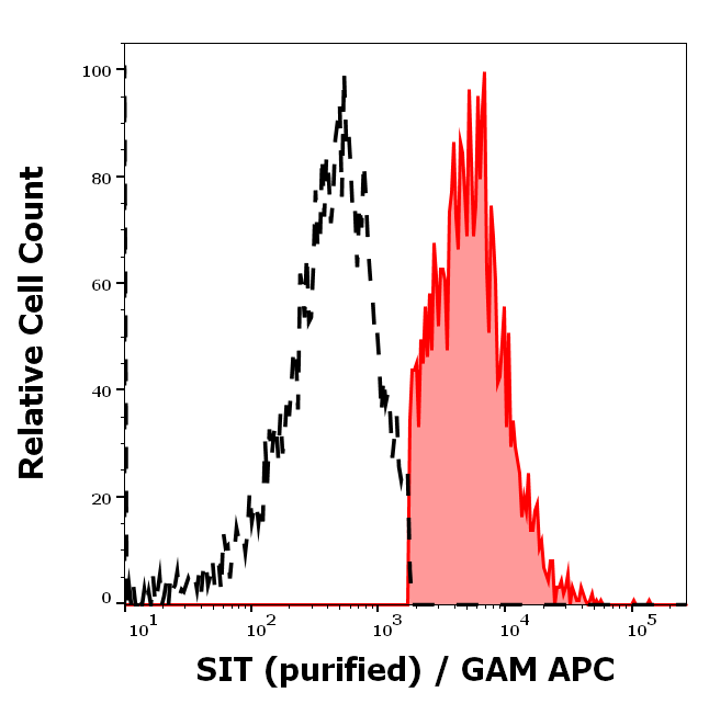
Separation of human CD3 negative SIT positive lymphocytes (red-filled) from CD3 negative SIT negative lymphocytes (black-dashed) in flow cytometry analysis (intracellular staining) of peripheral whole blood stained using anti-SIT (SIT-01) purified antibody (concentration in sample 9 µg/ml, GAM APC).
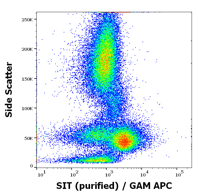
Flow cytometry intracellular staining pattern of human peripheral whole blood using anti-SIT (SIT-01) purified antibody (concentration in sample 9 µg/ml, GAM APC).
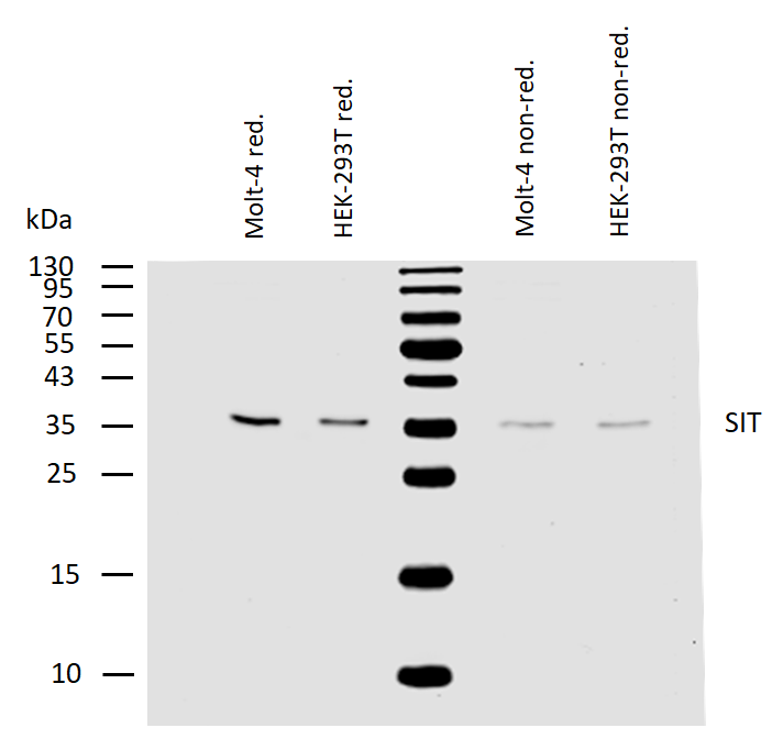
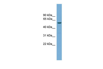
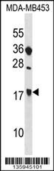
Western blotting analysis of human SIT using mouse monoclonal antibody SIT-01 on lysates of Molt-4 and HEK-293T cells under reducing and non-reducing conditions. Nitrocellulose membrane was probed with 2 µg/ml of mouse anti-SIT monoclonal antibody followed by IRDye800-conjugated anti-mouse secondary antibody. SIT was detected around 36 kDa.
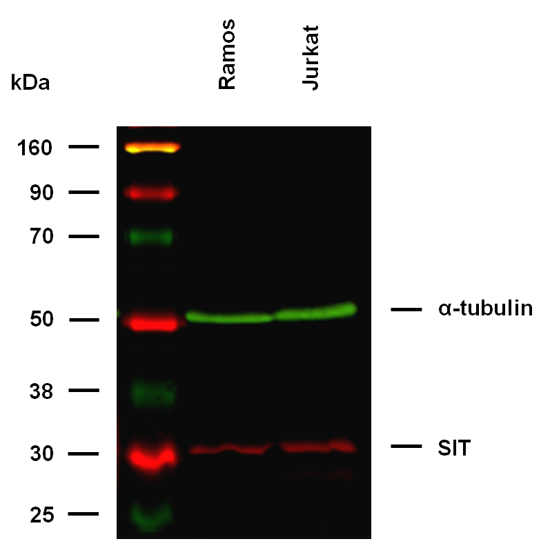
Anti-SIT Purified (clone SIT-01) works in WB application. Western blotting analysis was performed on whole cell extracts (RIPA lysis buffer) of Ramos and Jurkat cell lines, mixed and heated (100°C, 5 min) with reducing (2-mercaptoethanol) SDS-loading buffer. Samples were resolved using 10% Tris-glycine SDS gel electrophoresis. Nitrocellulose membrane blot was probed simultaneously with mouse IgG1 monoclonal antibody SIT-01 (2 µg/ml), and rat IgG2a anti-tubulin monoclonal antibody YOL1/34 (1 µg/ml) used as the loading control. Subclass-specific secondary antibodies IRDye 800CW Goat-anti-Rat IgG (green) and IRDye 680LT Goat-anti-Mouse IgG (red) were used for multiplex fluorescent Western blot detection. SIT was detected at ~32 kDa in tested cell lines.

Anti-SIT Purified (clone SIT-01) specificity verification by WB. The specificity of SIT-01 antibody was assessed by comparing binding signals in HEK293T cells overexpressing the target SIT protein to wild type cells (control) with low level of endogenous protein expression. Western blotting analysis was performed on whole cell extracts (urea lysis buffer) of transfected and control cells, mixed and heated (100°C, 5 min) with reducing (2-mercaptoethanol) SDS-loading buffer. Samples were resolved using 10% Tris-glycine SDS gel electrophoresis. Nitrocellulose membrane blot was probed simultaneously with mouse IgG1 monoclonal antibody SIT-01 (2 µg/ml), and rat IgG2a anti-tubulin monoclonal antibody YOL1/34 (1 µg/ml) used as the loading control. Subclass-specific secondary antibodies IRDye 800CW Goat-anti-Rat IgG (green) and IRDye 680LT Goat-anti-Mouse IgG (red) were used for multiplex fluorescent Western blot detection.
Documents Download
Datasheet
Product Information
Request a Document
Protocol Information
WB
Western Blot (IB, immunoblot)
FC
Flow Cytometry
IP
Immunoprecipitation
SIT Antibody (orb44488)

- 0.0
Based on 0 reviews
Participating in our Biorbyt product reviews program enables you to support fellow scientists by sharing your firsthand experience with our products.
Login to Submit a ReviewAvailable Sizes
Select a size below