You have no items in your shopping cart.
Description
Research Area
Cell Biology
Images & Validation
−Item 1 of 3
| Tested Applications | IF, IHC-P, WB |
|---|---|
| Dilution Range | IF - 1:50-100, IHC-P - 1:50-100 |
| Reactivity | Human, Mouse |
Key Properties
−| Antibody Type | Primary Antibody |
|---|---|
| Host | Rabbit |
| Clonality | Polyclonal |
| Isotype | Rabbit IgG |
| Immunogen | This BNIP3 antibody is generated from rabbits immunized with a KLH conjugated synthetic peptide between 215-252 amino acids from human BNIP3. Antigen Region: 215-252 aa. |
| Target | BNIP3 (HGNC:1084) |
| Molecular Weight | 21541 Da |
| Conjugation | Unconjugated |
Storage & Handling
−| Storage | Maintain refrigerated at 2-8°C for up to 2 weeks. For long term storage store at -20°C in small aliquots to prevent freeze-thaw cycles |
|---|---|
| Form/Appearance | Purified polyclonal antibody supplied in PBS with 0.09% (W/V) sodium azide. This antibody is prepared by Saturated Ammonium Sulfate (SAS) precipitation followed by dialysis against PBS. |
| Expiration Date | 12 months from date of receipt. |
| Disclaimer | For research use only |
Alternative Names
−BCL2/adenovirus E1B 19 kDa protein-interacting protein 3, BNIP3, NIP3
Similar Products
−Quality Guarantee
Explore bioreagents carefree to elevate your research. All our products are rigorously tested for performance. If a product does not perform as described on its datasheet, our scientific support team will provide expert troubleshooting, a prompt replacement, or a refund. For full details, please see our Terms & Conditions and Buying Guide. Contact us at support@biorbyt.com.
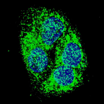
Fluorescent confocal image of HepG2 cells stained with BNIP3 (BH3 Domain Specific) Antibody. HepG2 cells were fixed with 4% PFA (20 min), permeabilized with Triton X-100 (0.2%, 30 min). Cells were then incubated with BNIP3 (BH3 Domain Specific) primary antibody (1:500, 2 h at room temperature). For secondary antibody, Alexa Fluor 488 conjugated donkey anti-rabbit antibody (green) was used (1:1000, 1h). Nuclei were counterstained with Hoechst 33342 (blue) (10 μg/ml, 5 min). BNIP3 immunoreactivity is localized to the cytoplasm of HepG2 cells.
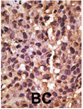
Formalin-fixed and paraffin-embedded human cancer tissue reacted with the primary antibody, which was peroxidase-conjugated to the secondary antibody, followed by DAB staining. This data demonstrates the use of this antibody for immunohistochemistry; clinical relevance has not been evaluated. BC = breast carcinoma; HC = hepatocarcinoma.
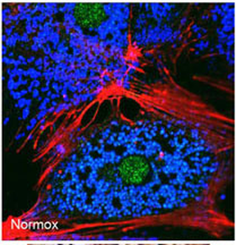
Freshly isolated mouse hepatocytes plated on coverslips (2 x10⁵ cells/22-mm glass coverslip) were cultured under normoxic conditions for 6 hr. The cells were then fixed in 2% paraformaldehyde in PBS for 1 hr, and processed for confocal immunofluorescence (red: F-actin, blue: ATP-synthase, green: BNIP3). Fluorescence labeling of BNIP3 accomplished with anti-BNIP3 Antibody.
Quick Database Links
UniProt Details
− No UniProt data available
NCBI Reference Sequences
−Associated Accession Numbers
Curated reference sequences for the gene transcript and protein product| Protein | NP_004043.2 |
|---|
Documents Download
Datasheet
Product Information
Request a Document
Protocol Information
WB
Western Blot (IB, immunoblot)
IHC-P
Immunohistochemistry Paraffin
IF
Immunofluorescence
BNIP3 Antibody (BH3 Domain Specific) (orb1936838)

- 0.0
Based on 0 reviews
Participating in our Biorbyt product reviews program enables you to support fellow scientists by sharing your firsthand experience with our products.
Login to Submit a ReviewAvailable Sizes
Select a size below
Choose Conjugation or Carrier Free Version
Free Secondary Antibody (20 ul)0/0
Please add an antibody product to your cart first.