You have no items in your shopping cart.
BrdU Antibody
SKU: orb345680
Description
Images & Validation
−Item 1 of 4
| Tested Applications | DOT, ELISA, FC, IF, IHC |
|---|---|
| Dilution Range | ELISA: 1:2000 - 1:10,000, FC: 1:50-1:100, IHC: 1:100-1:500, IF: 1:500 |
| Reactivity | Other |
| Application Notes |
Key Properties
−| Antibody Type | Primary Antibody |
|---|---|
| Host | Rabbit |
| Clonality | Polyclonal |
| Isotype | IgG |
| Immunogen | Anti-BrdU affinity purified antibody was purified from monospecific rabbit antiserum prepared via repeated immunizations with BromodeoxyUridine-KLH. |
| Purity | Anti-BrdU Antibody was affinity purified from monospecific antiserum by immunoaffinity chromatography. |
| Conjugation | Unconjugated |
Storage & Handling
−| Storage | Store BrdU Antibody at -20° C prior to opening. Aliquot contents and freeze at -20° C or below for extended storage. Avoid cycles of freezing and thawing. Centrifuge product if not completely clear after standing at room temperature. This product is stable for several weeks at 4° C as an undiluted liquid. Dilute only prior to immediate use. |
|---|---|
| Form/Appearance | Liquid (sterile filtered) |
| Buffer/Preservatives | Preservative: 0.01% (w/v) Sodium Azide. Stabilizer: None; Buffer: 0.02 M Potassium Phosphate, 0.15 M Sodium Chloride, pH 7.2 |
| Concentration | 1.23 mg/mL |
| Expiration Date | 12 months from date of receipt. |
| Dry Ice Shipping | Please note: This product requires shipment on dry ice. A dry ice surcharge will apply. |
| Disclaimer | For research use only |
Alternative Names
−rabbit anti-BrdU Antibody, bromodeoxyuridine, 5-bromo-2'-deoxyuridine
Similar Products
−- Item 1 of 6
Mouse Bromodeoxyuridine / BrdU Antibody [orb98204]
FC, ICC, IHC-Fr, IHC-P
Other
Mouse
Monoclonal
Unconjugated
0.1 mg - Item 1 of 3
BrdU (Proliferation Marker) Rabbit Polyclonal Antibody [orb10204]
ELISA
Human, Other
All
Rabbit
Polyclonal
Unconjugated
100 μl, 50 μl, 200 μl - Item 1 of 1
Mouse BrdU (bromodeoxyuridine), conjugated with FITC Antibody [orb108872]
FACS, IHC-P
Mouse
Monoclonal
FITC
100 μg - Item 1 of 1
- Item 1 of 3


Quality Guarantee
Explore bioreagents carefree to elevate your research. All our products are rigorously tested for performance. If a product does not perform as described on its datasheet, our scientific support team will provide expert troubleshooting, a prompt replacement, or a refund. For full details, please see our Terms & Conditions and Buying Guide. Contact us at support@biorbyt.com.
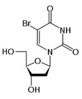
Bromodeoxyuridine (BrdU) chemical structure representation.
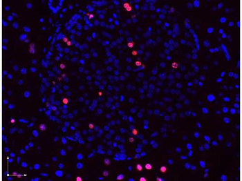
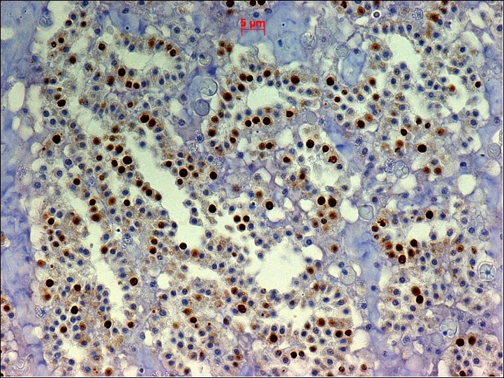
Immunofluorescence microscopy images of paraformaldehyde-fixed, paraffin-embedded pancreas sections stained with antibodies against BrdU (red or pink) and counterstained with DAPI (blue) and imaged with a 40× objective. DAPI stained nuclei (blue) indicate non-dividing cells, immunostained red and pink nuclei indicate actively dividing pancreatic ß-cells. The antibodies were diluted to 2.7 µg/ml. and incubated with tissue sections overnight at 4 degrees. Donkey anti-rabbit secondary antibody was diluted 1:2500.
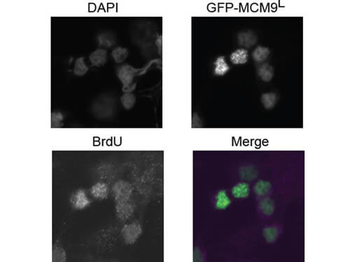
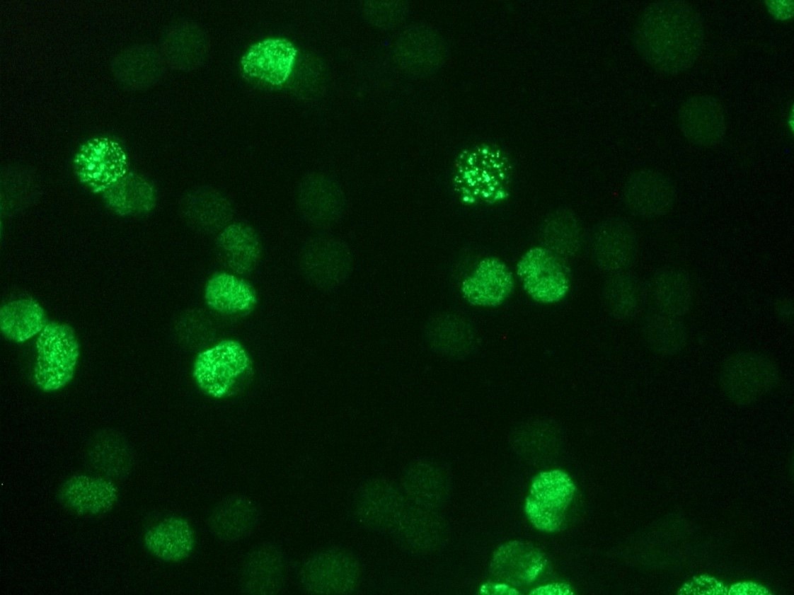
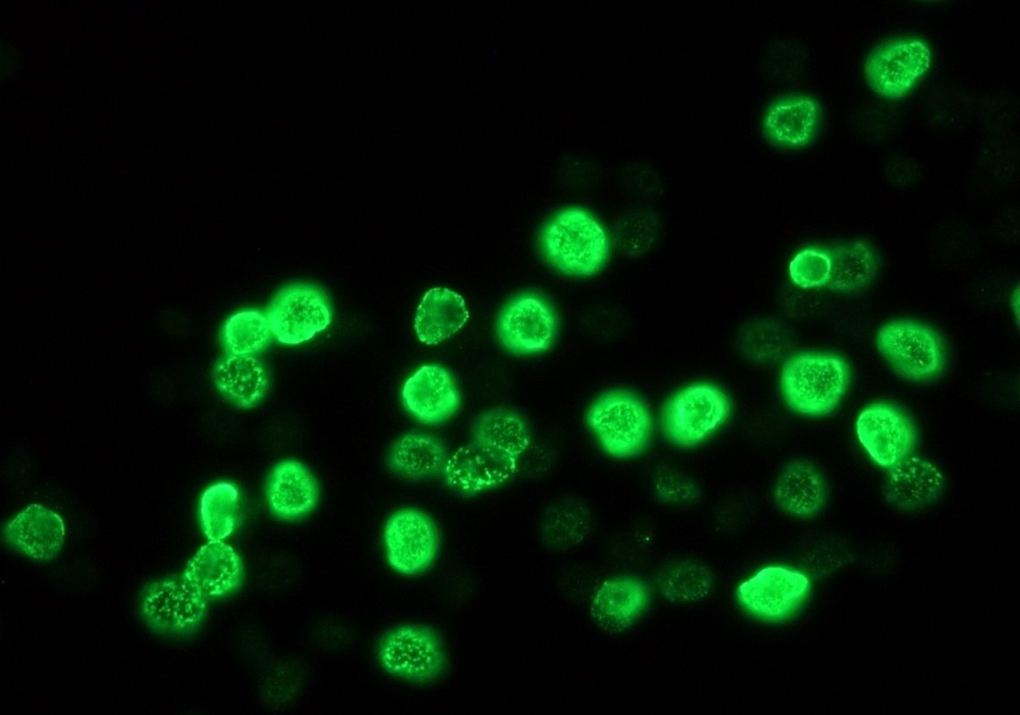
Immunofluorescence Microscopy of Rabbit anti-BrdU antibody. Tissue: 293T cells transfected expressing GFP-MCM9 L. Fixation: 0.5% PFA. Antigen retrieval: not required. Primary antibody: BrdU antibody at 10 µg/ml for 1 h at RT. Secondary antibody: Anti-rabbit ATTO550 secondary antibody at 1:10000 for 45 min at RT. Localization: BrdU is nuclear. Staining: BrdU in merged image shows with green and purple fluorescent signal with DAPI nuclear counterstain.
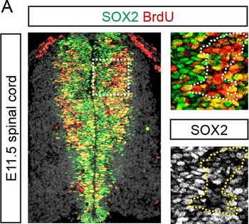
SOX2 represses proliferation in the developing spinal cord and stomach.(A) Percentage of cells expressing high or low levels of SOX2 labelled by a one hour pulse of BrdU in the E11.5 mouse spinal cord. Dotted lines in insets surround area of greatest BrdU incorporation. (B) Average background normalized SOX2 expression level and percentage of cells labelled by a one hour pulse of BrdU in the E11.5, E13.5 and E15.5 anterior and posterior stomach. (C) Percentage of electroporated cells in the chick spinal cord labelled by a 30 minute pulse of BrdU following misexpression of GFP, SOX2 or dnSOXB1. (D) Percentage of electroporated cells in E13.5 stomach explants labelled by a 30 minute pulse of BrdU following overexpression of GFP, SOX2 or dnSOXB1. All error bars represent standard deviations between experiments and p-values are calculated with two sided, unpaired t-tests (* = P < 0.05, ** = P < 0.01, *** = P < 0.001).
Documents Download
Datasheet
Product Information
Request a Document
Protocol Information
IHC
Immunohistochemistry
FC
Flow Cytometry
IF
Immunofluorescence
ELISA
Enzyme-linked Immunosorbent Assay (EIA)
DOT
Dot Blot
BrdU Antibody (orb345680)

- 0.0
Based on 0 reviews
Participating in our Biorbyt product reviews program enables you to support fellow scientists by sharing your firsthand experience with our products.
Login to Submit a ReviewAvailable Sizes
Select a size below