You have no items in your shopping cart.
Description
Research Area
Cancer Biology, Cell Biology, Immunology & Inflammation
Images & Validation
−Item 1 of 5
| Tested Applications | IF, IHC-P, WB |
|---|---|
| Dilution Range | IF - 1:25, WB - 1:2000, IHC-P - 1:25 |
| Reactivity | Human, Mouse |
Key Properties
−| Antibody Type | Primary Antibody |
|---|---|
| Host | Rabbit |
| Clonality | Polyclonal |
| Isotype | Rabbit IgG |
| Immunogen | This CDKN1A antibody is generated from a rabbit immunized with a KLH conjugated synthetic peptide between 133-164 amino acids from the C-terminal region of human CDKN1A. |
| Target | CDKN1A (HGNC:1784) |
| Molecular Weight | 18119 Da |
| Conjugation | Unconjugated |
Storage & Handling
−| Storage | Maintain refrigerated at 2-8°C for up to 2 weeks. For long term storage store at -20°C in small aliquots to prevent freeze-thaw cycles |
|---|---|
| Form/Appearance | Purified polyclonal antibody supplied in PBS with 0.09% (W/V) sodium azide. This antibody is purified through a protein A column, followed by peptide affinity purification. |
| Expiration Date | 12 months from date of receipt. |
| Disclaimer | For research use only |
Alternative Names
−Cyclin-dependent kinase inhibitor 1, CDK-interacting protein 1, Melanoma differentiation-associated protein 6, MDA-6, p21, CDKN1A, CAP20, CDKN1, CIP1, MDA6, PIC1, SDI1, WAF1
Similar Products
−- Item 1 of 4
p21 (CDKN1A) Antibody (C-term) [orb38089]
FC, IF, IHC-P, WB
Human
Rabbit
Polyclonal
Unconjugated
50 μl, 100 μl - Item 1 of 4
CDKN1A Antibody (C-term) [orb1936665]
IF, IHC-P, WB
Human, Mouse
Rabbit
Polyclonal
Unconjugated
50 μl, 100 μl - Item 1 of 1
- Item 1 of 1
Quality Guarantee
Explore bioreagents carefree to elevate your research. All our products are rigorously tested for performance. If a product does not perform as described on its datasheet, our scientific support team will provide expert troubleshooting, a prompt replacement, or a refund. For full details, please see our Terms & Conditions and Buying Guide. Contact us at support@biorbyt.com.
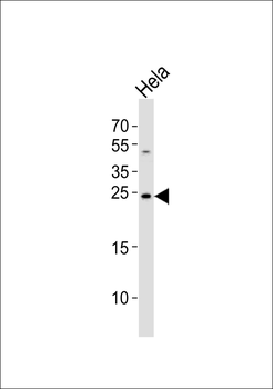
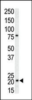
Western blot analysis of lysate from Hela cell line, using CDKN1A Antibody (C-term). Diluted at 1:1000. A goat anti-rabbit IgG H&L (HRP) at 1:10000 dilution was used as the secondary antibody. Lysate at 35 ug.
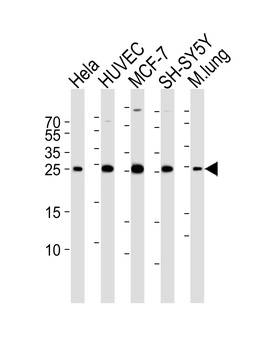
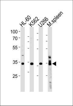
Western blot analysis of lysates from Hela, HUVEC, MCF-7, SH-SY5Y cell line and mouse lung tissue lysate (from left to right), using CDKN1A Antibody (C-term). Diluted at 1:1000 at each lane. A goat anti-rabbit IgG H&L (HRP) at 1:5000 dilution was used as the secondary antibody. Lysates at 35 ug per lane.
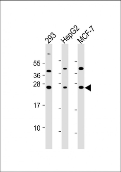
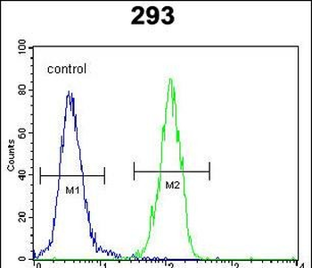
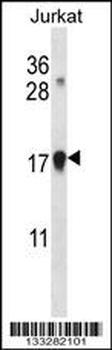
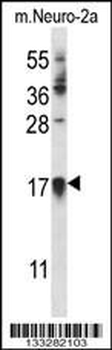
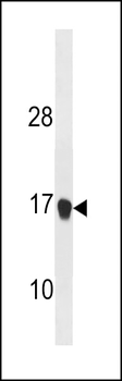
All lanes: Anti-CDKN1A Antibody (C-term) at 1:2000 dilution. Lane 1: 293 whole cell lysate. Lane 2: HepG2 whole cell lysate. Lane 3: MCF-7 whole cell lysate. Lysates/proteins at 20 µg per lane. Secondary Goat Anti-Rabbit IgG, (H+L), Peroxidase conjugated at 1/10000 dilution. Predicted band size: 18 kDa. Blocking/Dilution buffer: 5% NFDM/TBST.
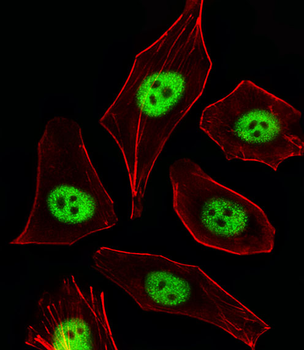
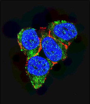
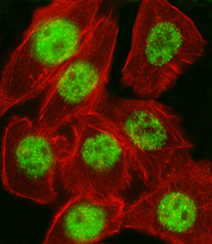
Fluorescent image of A549 cells stained with CDKN1A Antibody (C-term). Diluted at 1:25 dilution. An Alexa Fluor 488-conjugated goat anti-rabbit lgG at 1:400 dilution was used as the secondary antibody (green). Cytoplasmic actin was counterstained with Alexa Fluor 555 conjugated with Phalloidin (red). Cytoplasm
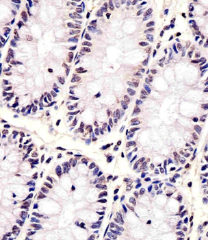
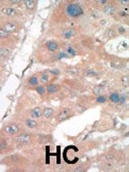
Staining CDKN1A in human colon tissue sections by Immunohistochemistry (IHC-P - paraformaldehyde-fixed, paraffin-embedded sections). Tissue was fixed with formaldehyde and blocked with 3% BSA for 0.5 hour at room temperature; antigen retrieval was by heat mediation with a citrate buffer (pH6). Samples were incubated with primary antibody (1/25) for 1 hours at 37°C. A undiluted biotinylated goat polyvalent antibody was used as the secondary antibody.
Quick Database Links
Gene Symbol
CDKN1A (HGNC:1784)
UniProt
UniProt Details
− No UniProt data available
Documents Download
Datasheet
Product Information
Request a Document
Protocol Information
WB
Western Blot (IB, immunoblot)
IHC-P
Immunohistochemistry Paraffin
IF
Immunofluorescence
CDKN1A Antibody (C-term) (orb1927120)

- 0.0
Based on 0 reviews
Participating in our Biorbyt product reviews program enables you to support fellow scientists by sharing your firsthand experience with our products.
Login to Submit a ReviewAvailable Sizes
Select a size below
Choose Conjugation or Carrier Free Version
Free Secondary Antibody (20 ul)0/0
Please add an antibody product to your cart first.