You have no items in your shopping cart.
Collagen Type III Antibody Peroxidase Conjugated
SKU: orb345846
Description
Images & Validation
−Item 1 of 1
| Tested Applications | ELISA, IHC, IP, WB |
|---|---|
| Dilution Range | ELISA: 1:10,000 - 1:50,000, IHC: 1:200 - 1:1,000, IP: 1:100, WB: 1:1,000 - 1:5,000 |
| Reactivity | Bovine, Human |
| Application Notes |
Key Properties
−| Antibody Type | Primary Antibody |
|---|---|
| Host | Rabbit |
| Clonality | Polyclonal |
| Isotype | IgG |
| Immunogen | Collagen Type III from human and bovine placenta |
| Purity | This product has been prepared by immunoaffinity chromatography using immobilized antigens. Some class-specific anti-collagens may be specific for three-dimensional epitopes which may result in diminished reactivity with denatured collagen or formalin-fixed, paraffin embedded tissues. This antibody reacts with most mammalian Type III collagens and has expected cross-reactivity with Type I and negligible cross reactivity with Type II, IV, V or VI collagens. Non-specific cross-reaction of anti-collagen antibodies with other human serum proteins or non-collagen extracellular matrix proteins has not been tested. |
| Conjugation | HRP |
Storage & Handling
−| Storage | Store vial at 4° C prior to restoration. For extended storage aliquot contents and freeze at -20° C or below. Avoid cycles of freezing and thawing. Centrifuge product if not completely clear after standing at room temperature. This product is stable for several weeks at 4° C as an undiluted liquid. Dilute only prior to immediate use. |
|---|---|
| Form/Appearance | Lyophilized |
| Buffer/Preservatives | Preservative: 0.01% (w/v) Thimerosal. Stabilizer: 10 mg/mL Bovine Serum Albumin (rAlbumin) - Immunoglobulin and Protease free; Buffer: 0.01 M Sodium Phosphate, 0.25 M Sodium Chloride, pH 7.2 |
| Concentration | 1.0 mg/mL |
| Expiration Date | 12 months from date of receipt. |
| Disclaimer | For research use only |
Alternative Names
−rabbit anti-Collagen Type III antibody peroxidase conjugation, HRP conjugated rabbit anti-Collagen Type III antibody, Collagen type III alpha 1 antibody, Collagen type III alpha antibody, EDS4A antibody, Ehlers Danlos syndrome type IV, autosomal dominant antibody, Fetal collagen antibody, COL3A1, Collagen alpha-1 (III) chain
Similar Products
−- Item 1 of 1
Quality Guarantee
Explore bioreagents carefree to elevate your research. All our products are rigorously tested for performance. If a product does not perform as described on its datasheet, our scientific support team will provide expert troubleshooting, a prompt replacement, or a refund. For full details, please see our Terms & Conditions and Buying Guide. Contact us at support@biorbyt.com.
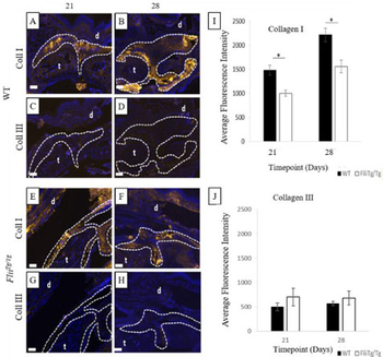
Immunohistochemistry of Anti-Collagen Type III Peroxidase Conjugated. WT mice have significantly increased collagen type I expression in the tendon adhesions. Representative images of collagen type I expression (gold) in the tendon adhesions of WT (a-b) and FliiTg/Tg (e-f) mice and collagen type III expression (gold) in the tendon adhesions of WT (c-d) FliiTg/Tg (g-h) at 21 and 28 days post 50% partial laceration injury. (i) WT mice have significantly upregulated collagen type I levels in the adhesions compared with FliiTg/Tg mice. (j) No significant difference was noted in collagen type III levels in WT and FliiTg/Tg mice, although detectable levels of collagen type III were found in the FliiTg/Tg mice. DAPI is represented as blue fluorescence; t, tendon; d, dermis. Dotted line represents tendon adhesion area. Magnification × 10. Scale bar = 200 µm and refers to all images. Data represented as mean ± SEM. *p ≤ 0.05. n = 6.
Quick Database Links
UniProt
RefSeq (Protein):NP_000081.1
UniProt Details
− No UniProt data available
NCBI Reference Sequences
−Associated Accession Numbers
Curated reference sequences for the gene transcript and protein product| Protein | NP_000081.1 |
|---|
Documents Download
Datasheet
Product Information
Request a Document
Protocol Information
WB
Western Blot (IB, immunoblot)
IHC
Immunohistochemistry
ELISA
Enzyme-linked Immunosorbent Assay (EIA)
IP
Immunoprecipitation
Collagen Type III Antibody Peroxidase Conjugated (orb345846)

- 0.0
Based on 0 reviews
Participating in our Biorbyt product reviews program enables you to support fellow scientists by sharing your firsthand experience with our products.
Login to Submit a ReviewAvailable Sizes
Select a size below