You have no items in your shopping cart.
Description
Images & Validation
−Item 1 of 2
| Tested Applications | DOT, ELISA, FC, IF, WB |
|---|---|
| Dilution Range | ELISA: 1:20,000 - 1:40,000, FC: 1:2000, IF: 1:500 - 1:2,500, WB: 1:10,000-1:25,000 |
| Reactivity | Other |
| Application Notes |
Key Properties
−| Antibody Type | Primary Antibody |
|---|---|
| Host | Rabbit |
| Clonality | Polyclonal |
| Isotype | IgG |
| Immunogen | Anti-Green Fluorescent Protein (GFP) is produced by immunizing GFP containing fusion protein that corresponds to the full length amino acid sequence (246aa) derived from the jellyfish Aequorea victoria. |
| Purity | GFP Antibody Fluorescein Conjugated was prepared from monospecific antiserum by immunoaffinity chromatography using Green Fluorescent Protein (Aequorea victoria) coupled to agarose beads followed by solid phase adsorption(s) to remove any unwanted reactivities. Assay by immunoelectrophoresis resulted in a single precipitin arc against anti-Rabbit Serum, anti-Fluorescein and purified and partially purified Green Fluorescent Protein (Aequorea victoria) Serum. No reaction was observed against Human, Mouse and Rat Serum Proteins. |
| Conjugation | FITC |
Storage & Handling
−| Storage | Store vial at 4° C prior to restoration. For extended storage aliquot contents and freeze at -20° C or below. Avoid cycles of freezing and thawing. Centrifuge product if not completely clear after standing at room temperature. This product is stable for several weeks at 4° C as an undiluted liquid. Dilute only prior to immediate use. |
|---|---|
| Form/Appearance | Lyophilized |
| Buffer/Preservatives | Preservative: 0.01% (w/v) Sodium Azide. Stabilizer: 10 mg/mL Bovine Serum Albumin (rAlbumin) - Immunoglobulin and Protease free; Buffer: 0.02 M Potassium Phosphate, 0.15 M Sodium Chloride, pH 7.2 |
| Concentration | 1.0 mg/mL |
| Expiration Date | 12 months from date of receipt. |
| Disclaimer | For research use only |
Alternative Names
−rabbit anti-GFP antibody fluorescein conjugation, FITC conjugated rabbit anti-GFP antibody, Green Fluorescent Protein, GFP antibody, Green Fluorescent Protein antibody, EGFP, enhanced Green Fluorescent Protein, Aequorea victoria, Jellyfish
Similar Products
−- Item 1 of 2
- Item 1 of 1

Quality Guarantee
Explore bioreagents carefree to elevate your research. All our products are rigorously tested for performance. If a product does not perform as described on its datasheet, our scientific support team will provide expert troubleshooting, a prompt replacement, or a refund. For full details, please see our Terms & Conditions and Buying Guide. Contact us at support@biorbyt.com.

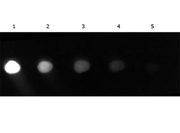
Biorbyt FITC conjugated Rabbit anti GFP (green) stains mouse spleen cells Tissue: spleen cells infected with MHV68-H2bYFP.

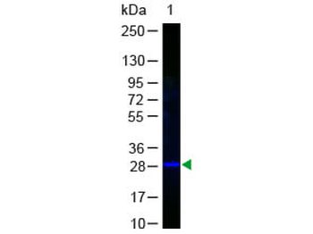
Western Blot of GFP Antibody Fluorescein Conjugated. Lane 1: GFP. Load: 50 ng per lane. Primary antibody: none. Secondary antibody: Fluorescein Conjugated Anti-GFP at 1:1000 for 60 min at RT. Block: 1% BSA-TTBS for 30 min at RT. Predicted/Observed size: 28 kDa, 28 kDa.
Quick Database Links
UniProt
UniProt Details
− No UniProt data available
Documents Download
Datasheet
Product Information
Request a Document
Protocol Information
WB
Western Blot (IB, immunoblot)
FC
Flow Cytometry
IF
Immunofluorescence
ELISA
Enzyme-linked Immunosorbent Assay (EIA)
DOT
Dot Blot
GFP Antibody Fluorescein Conjugated (orb345838)

- 0.0
Based on 0 reviews
Participating in our Biorbyt product reviews program enables you to support fellow scientists by sharing your firsthand experience with our products.
Login to Submit a ReviewAvailable Sizes
Select a size below
Choose Conjugation or Carrier Free Version
Free Secondary Antibody (20 ul)0/0
Please add an antibody product to your cart first.