You have no items in your shopping cart.
Description
Research Area
Cancer Biology, Cell Biology, Protein Biochemistry, Signal Transduction
Images & Validation
−Item 1 of 3
| Tested Applications | ELISA, ICC, IF, IHC, WB |
|---|---|
| Dilution Range | WB (1:1000); ELISA (1:1000); ICC/IF (1:60); IHC (1:50) |
| Reactivity | Human, Mouse, Rat |
| Application Notes |
Key Properties
−| Host | Rabbit |
|---|---|
| Clonality | Polyclonal |
| Immunogen | Synthetic peptide from the N-terminal of Human HSP70 Acetyl Lys77 (1-100 aa), conjugated to Keyhole Limpet Haemocyanin (KLH). |
| Target | HSP70 (Acetyl Lys77) |
| Molecular Weight | 70 kDa |
| Purification | Peptide Affinity Purified |
| Conjugation | FITC |
Storage & Handling
−| Storage | Conjugated antibodies should be stored according to the product label |
|---|---|
| Buffer/Preservatives | 640.91mM DMSO, 136.36 mM Ethanolamine, 126.89 mM chlorides, 9.09mM phosphates, 9.09mM NaHCO3 |
| Concentration | 0.5mg/mL |
| Expiration Date | 12 months from date of receipt. |
| Disclaimer | For research use only |
Alternative Names
−HSPA1A, HSPA1B, HSPA1, HSP70, HSP70-1, HSP70.1, HSP70-2, HSP72, HSP73, HSX70, Heat shock 70 kDa protein 1A, Heat shock 70 kDa protein 1B, HSP70 Acetylated lysine 77, HSP70 Acetyl Lys77
Quality Guarantee
Explore bioreagents carefree to elevate your research. All our products are rigorously tested for performance. If a product does not perform as described on its datasheet, our scientific support team will provide expert troubleshooting, a prompt replacement, or a refund. For full details, please see our Terms & Conditions and Buying Guide. Contact us at support@biorbyt.com.
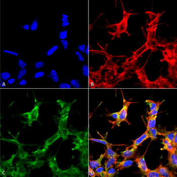
Immunocytochemistry/Immunofluorescence analysis using Rabbit Anti-HSP70 Acetyl Lys77 Polyclonal Antibody. Tissue: Embryonic kidney cells (HEK293) cultured overnight with 50 μM H2O2. Species: Human. Fixation: 5% Formaldehyde for 5 min. Primary Antibody: Rabbit Anti-HSP70 Acetyl Lys77 Polyclonal Antibody at 1:60 for 30-60 min at RT. Secondary Antibody: Goat Anti-Rabbit Alexa Fluor 488 at 1:1500 for 30-60 min at RT. Counterstain: Phalloidin Alexa Fluor 633 F-Actin stain; DAPI (blue) nuclear stain at 1:250, 1:50000 for 30-60 min at RT. Localization: Cytoplasmic. Magnification: 20X (2X Zoom). (A) DAPI (blue) nuclear stain. (B) Phalloidin Alex Fluor 633 F-Actin stain. (C) HSP70 Antibody (Acetyl Lys77). (D) Composite.

Immunohistochemistry analysis using Rabbit Anti-HSP70 (Acetyl Lys77) Polyclonal Antibody. Tissue: Colon Cancer. Species: Human. Fixation: Formalin Fixed Paraffin-Embedded. Primary Antibody: Rabbit Anti-HSP70 (Acetyl Lys77) Polyclonal Antibody at 1:50 for 30 min at RT. Counterstain: Hematoxylin. Magnification: 20X. HRP-DAB Detection.
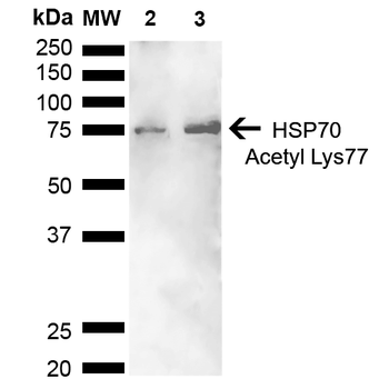
Western blot analysis of Human Cervical cancer cell line (HeLa) lysate showing detection of ~70 kDa HSP70 Acetyl Lys77 protein using Rabbit Anti-HSP70 Acetyl Lys77 Polyclonal Antibody. Lane 1: Molecular Weight Ladder (MW). Lane 2: Cervical Cancer cell line (HeLa) lysate. Lane 3: H2O2 Cervical Cancer cell line (HeLa) lysate. Load: 10 μg. Block: 5% Skim Milk in 1X TBST. Primary Antibody: Rabbit Anti-HSP70 Acetyl Lys77 Polyclonal Antibody at 1:1000 for 2 hours at RT. Secondary Antibody: Goat Anti-Rabbit HRP:IgG at 1:3000 for 1 hour at RT. Color Development: ECL solution for 5 min at RT. Predicted/Observed Size: ~70 kDa.
Quick Database Links
UniProt Details
− No UniProt data available
NCBI Gene Details
− No NCBI Gene data available
NCBI Reference Sequences
−Associated Accession Numbers
Curated reference sequences for the gene transcript and protein product| Protein | NP_005336.3 |
|---|
Documents Download
Datasheet
Product Information
Request a Document
Protocol Information
WB
Western Blot (IB, immunoblot)
IHC
Immunohistochemistry
IF
Immunofluorescence
ICC
Immunocytochemistry
ELISA
Enzyme-linked Immunosorbent Assay (EIA)
HSP70 (Acetyl Lys77) Antibody (FITC) (orb414155)

- 0.0
Based on 0 reviews
Participating in our Biorbyt product reviews program enables you to support fellow scientists by sharing your firsthand experience with our products.
Login to Submit a ReviewAvailable Sizes
Select a size below
Choose Conjugation or Carrier Free Version
Free Secondary Antibody (20 ul)0/0
Please add an antibody product to your cart first.