You have no items in your shopping cart.
Description
Research Area
Pharmacology & Drug Discovery
Images & Validation
−Item 1 of 9
| Tested Applications | ELISA, IP, WB |
|---|---|
| Dilution Range | ELISA: 1:1,000-1:2,500, IP: 10 µg/mg protein sample, WB: 1:500-1:1,000 |
| Application Notes |
Key Properties
−| Antibody Type | Primary Antibody |
|---|---|
| Host | Rabbit |
| Clonality | Polyclonal |
| Isotype | IgG |
| Immunogen | Anti-Lysine Acetylated Antibody was prepared from whole rabbit serum produced by repeated immunizations with acetylated lysine conjugated KLH. |
| Purity | Anti-Lysine Acetylated Antibody was prepared from monospecific antiserum by immunoaffinity chromatography using acetylated lysine peptide coupled to agarose. Assay by immunoelectrophoresis resulted in a single precipitin arc against anti-Rabbit IgG. The antibody reacts specifically with acetylated lysine residues. |
| Conjugation | Unconjugated |
Storage & Handling
−| Storage | Store vial at -20° C prior to opening. Aliquot contents and freeze at -20° C or below for extended storage. Avoid cycles of freezing and thawing. Centrifuge product if not completely clear after standing at room temperature. This product is stable for several weeks at 4° C as an undiluted liquid. Dilute only prior to immediate use. |
|---|---|
| Form/Appearance | Liquid (sterile filtered) |
| Buffer/Preservatives | Preservative: None. Stabilizer: 50% (v/v) Glycerol; Buffer: 0.02 M Potassium Phosphate, 0.15 M Sodium Chloride, pH 7.2 |
| Concentration | 1.0 mg/ml |
| Expiration Date | 12 months from date of receipt. |
| Dry Ice Shipping | Please note: This product requires shipment on dry ice. A dry ice surcharge will apply. |
| Disclaimer | For research use only |
Alternative Names
−acetyl Lysine antibody, Acetylated lysine antibody, Lysine antibody
Similar Products
−- Item 1 of 2
Anti-H4 K8Ac; K12Ac; K16Ac [KM-2] [orb758977]
ELISA, IF, IHC, WB
Human
Mouse
Monoclonal
Unconjugated
0.2 mg - Item 1 of 2
Anti-H4 K8Ac; K12Ac; K16Ac [KM-2] [orb758978]
ELISA, IF, IHC, WB
Human
Mouse
Monoclonal
Unconjugated
0.2 mg - Item 1 of 4
- Item 1 of 4
- Item 1 of 4
Acetylated Lysine Antibody (PerCP) [orb182156]
ELISA, ICC, IF, IP, WB
All
Rabbit
Polyclonal
PerCP
100 μg
![Anti-H4 K8Ac; K12Ac; K16Ac [KM-2]](/images/pub/media/catalog/product/NewWebsite/35/orb758977_1.png)
![Anti-H4 K8Ac; K12Ac; K16Ac [KM-2]](/images/pub/media/catalog/product/NewWebsite/35/orb758977_2.png)
![Anti-H4 K8Ac; K12Ac; K16Ac [KM-2]](/images/pub/media/catalog/product/NewWebsite/35/orb758978_1.png)
![Anti-H4 K8Ac; K12Ac; K16Ac [KM-2]](/images/pub/media/catalog/product/NewWebsite/35/orb758978_2.png)


Quality Guarantee
Explore bioreagents carefree to elevate your research. All our products are rigorously tested for performance. If a product does not perform as described on its datasheet, our scientific support team will provide expert troubleshooting, a prompt replacement, or a refund. For full details, please see our Terms & Conditions and Buying Guide. Contact us at support@biorbyt.com.
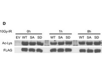
Acetylation does not crosstalk with IR-induced pSer824 and does not affect the co-repressor activity of KAP1.(A) Control and SIRT1 depleted HEK293T cells were harvested at indicated time points following 10Gy-IR. IR-induced KAP1 pSer824 was examined by phosphorylation specific antibody. (B) Cells transfected with FLAG-tagged WT-KAP1 or 4KR mutant were harvested at indicated time points following 4Gy-IR. Exogenous KAP1 was immunoprecipitated and pSer824 was examined by phosphorylation specific antibody. (C) Cyclin D3 luciferase construct was co-transfected with combinations of HA-tagged E2F1, FLAG-tagged KAP1, and 4KR mutant. Luciferase activity was measured 48 hours post transfection. (D) Cells transfected with FLAG-tagged WT-KAP1 or KAP1 phospho mutants (S824A and S824D) were harvested at indicated time points following 10Gy-IR. Recombinant KAP1 was purified by immunoprecipitation and the total acetylation level was assessed by immunoblotting.
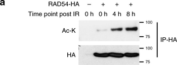
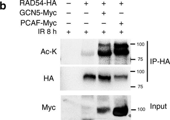
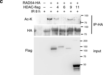
RAD54 acetylation is important for BRD9 recognition and HR activity. a–c RAD54 is acetylated by GCN5/PCAF and deacetylated by HDAC 6/HDAC11 following induction of DNA damage. a 293T cells were transfected with control or RAD54-HA plasmid. Twenty-four hours after transfection, cells were exposed to the 10-Gy IR and harvested at the indicated time points. Immunoprecipitation with anti-HA beads was performed. Blots were probed with the indicated antibodies. b 293T cells were transfected with the indicated plasmids. Twenty-four hours after transfection, cells were exposed to 10-Gy IR, and lysates were collected after 8 h. Immunoprecipitation with anti-HA beads was performed. Blots were probed with the indicated antibodies. c 293T cells were transfected with the indicated plasmids, treated as indicated, and subjected to immunoprecipitation as outlined in b. Blots were probed with the indicated antibodies. d, e RAD54 K515 acetylation is important for RAD51–RAD54 complex formation. d 293T cells were transfected with the indicated plasmids, treated as indicated, and subjected to immunoprecipitation as outlined in b. Blots were probed with the indicated antibodies. e 293T cells were transfected with the indicated plasmids, exposed to either no IR or 10-Gy IR as indicated, and subjected to immunoprecipitation as outlined in b. Blots were probed with the indicated antibodies. f–h RAD54 K515 acetylation is essential for HR activity. f, g U2OS cells were transfected with the indicated plasmids and exposed to 2-Gy IR. Cells were fixed after 8 h and stained for the indicated proteins. Representative immunofluorescence images of RAD51 (green) and RAD54 (red) are shown in f. Quantification of the indicated foci is shown in g. Representative data (mean ± SEM) are shown from n = 50 cells examined over three independent experiments. **p < 0.01, ***p < 0.001 by two-sided unpaired t test. NS not significant. Scale bar, 10 µm. h Survival curves of U2OS cells expressing the indicated constructs and exposed to the indicated doses of PARPi or cisplatin. Representative data (mean ± SEM) are shown from n = 3 biologically independent samples. *p < 0.05, ***p < 0.001 by two-sided unpaired t test.
RAD54 acetylation is important for BRD9 recognition and HR activity. a–c RAD54 is acetylated by GCN5/PCAF and deacetylated by HDAC 6/HDAC11 following induction of DNA damage. a 293T cells were transfected with control or RAD54-HA plasmid. Twenty-four hours after transfection, cells were exposed to the 10-Gy IR and harvested at the indicated time points. Immunoprecipitation with anti-HA beads was performed. Blots were probed with the indicated antibodies. b 293T cells were transfected with the indicated plasmids. Twenty-four hours after transfection, cells were exposed to 10-Gy IR, and lysates were collected after 8 h. Immunoprecipitation with anti-HA beads was performed. Blots were probed with the indicated antibodies. c 293T cells were transfected with the indicated plasmids, treated as indicated, and subjected to immunoprecipitation as outlined in b. Blots were probed with the indicated antibodies. d, e RAD54 K515 acetylation is important for RAD51–RAD54 complex formation. d 293T cells were transfected with the indicated plasmids, treated as indicated, and subjected to immunoprecipitation as outlined in b. Blots were probed with the indicated antibodies. e 293T cells were transfected with the indicated plasmids, exposed to either no IR or 10-Gy IR as indicated, and subjected to immunoprecipitation as outlined in b. Blots were probed with the indicated antibodies. f–h RAD54 K515 acetylation is essential for HR activity. f, g U2OS cells were transfected with the indicated plasmids and exposed to 2-Gy IR. Cells were fixed after 8 h and stained for the indicated proteins. Representative immunofluorescence images of RAD51 (green) and RAD54 (red) are shown in f. Quantification of the indicated foci is shown in g. Representative data (mean ± SEM) are shown from n = 50 cells examined over three independent experiments. **p < 0.01, ***p < 0.001 by two-sided unpaired t test. NS not significant. Scale bar, 10 µm. h Survival curves of U2OS cells expressing the indicated constructs and exposed to the indicated doses of PARPi or cisplatin. Representative data (mean ± SEM) are shown from n = 3 biologically independent samples. *p < 0.05, ***p < 0.001 by two-sided unpaired t test.
RAD54 acetylation is important for BRD9 recognition and HR activity. a–c RAD54 is acetylated by GCN5/PCAF and deacetylated by HDAC 6/HDAC11 following induction of DNA damage. a 293T cells were transfected with control or RAD54-HA plasmid. Twenty-four hours after transfection, cells were exposed to the 10-Gy IR and harvested at the indicated time points. Immunoprecipitation with anti-HA beads was performed. Blots were probed with the indicated antibodies. b 293T cells were transfected with the indicated plasmids. Twenty-four hours after transfection, cells were exposed to 10-Gy IR, and lysates were collected after 8 h. Immunoprecipitation with anti-HA beads was performed. Blots were probed with the indicated antibodies. c 293T cells were transfected with the indicated plasmids, treated as indicated, and subjected to immunoprecipitation as outlined in b. Blots were probed with the indicated antibodies. d, e RAD54 K515 acetylation is important for RAD51–RAD54 complex formation. d 293T cells were transfected with the indicated plasmids, treated as indicated, and subjected to immunoprecipitation as outlined in b. Blots were probed with the indicated antibodies. e 293T cells were transfected with the indicated plasmids, exposed to either no IR or 10-Gy IR as indicated, and subjected to immunoprecipitation as outlined in b. Blots were probed with the indicated antibodies. f–h RAD54 K515 acetylation is essential for HR activity. f, g U2OS cells were transfected with the indicated plasmids and exposed to 2-Gy IR. Cells were fixed after 8 h and stained for the indicated proteins. Representative immunofluorescence images of RAD51 (green) and RAD54 (red) are shown in f. Quantification of the indicated foci is shown in g. Representative data (mean ± SEM) are shown from n = 50 cells examined over three independent experiments. **p < 0.01, ***p < 0.001 by two-sided unpaired t test. NS not significant. Scale bar, 10 µm. h Survival curves of U2OS cells expressing the indicated constructs and exposed to the indicated doses of PARPi or cisplatin. Representative data (mean ± SEM) are shown from n = 3 biologically independent samples. *p < 0.05, ***p < 0.001 by two-sided unpaired t test.
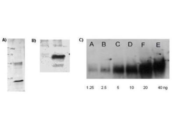
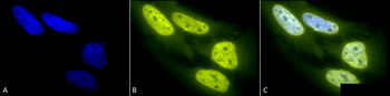
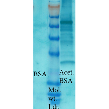
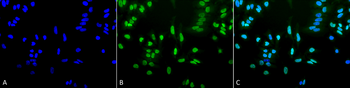
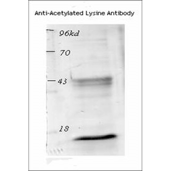
Biorbyt's Affinity Purified anti-Acetylated Lysine (AcK) antibody is shown to detect acetylated histone in TSA-treated mouse spleen cell lysate (Panel A); control (left lane) and TSA-treated mouse spleen cell lysate (right lane) in panel B; and in acetylated BSA loaded as indicated (panel C).
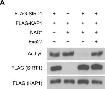
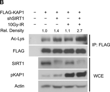
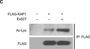
SIRT1 deacetylates KAP1 in vitro and in vivo.(A) SIRT1 deacetylates KAP1 in vitro. Exogenous FLAG-tagged KAP1 and SIRT1 were purified by anti-FLAG immunoprecipitation. Combination of purified proteins was incubated in deacetylation buffer supplemented with or without NAD+ cofactor. Ex527 (40 µm) was added to block the deacetylase activity of SIRT1. (B) SIRT1 deacetylates KAP1 in vivo. FLAG-tagged KAP1 was transfected into control or SIRT1 depleted HEK293T cells. Cells were treated with or without IR 1 hour before harvest. Cell lysates were subjected to immunoprecipitation using anti-FLAG antibody, followed by western blot analysis to assess the total KAP1 acetylation level. Relative density of the overall acetyl lysine was quantified using ImageJ software. Acetylation level was normalized to corresponding FLAG band. (C) HEK293T cells transfected with FLAG-tagged KAP1 were treated with DMSO or Ex527 (20 µm) for 6 hours before harvest. Cell lysates were then immunoprecipitated with FLAG-conjugated agarose beads, and the immunoprecipitates were blotted with anti-acetyl lysine antibody to determine the total acetylation level. (D and E) HEK293T cells were transfected with FLAG-tagged KAP1 and treated with DMSO or Ex527 (20 µm) for 6 hours before harvest. Recombinant KAP1 was purified and sent for mass spectrometric analysis. Acetyl residues with > 2-fold enhancement after inhibitor treatment were considered to be SIRT1 targeted sites. (F) Site-directed mutagenesis was applied to generate 4KR mutant. FLAG-tagged WT-KAP1 or 4KR mutant were transfected into control or SIRT1 depleted HEK293T cells. Total acetylation levels of recombinant WT-KAP1 and 4KR mutant were assessed by immunoblotting.
SIRT1 deacetylates KAP1 in vitro and in vivo.(A) SIRT1 deacetylates KAP1 in vitro. Exogenous FLAG-tagged KAP1 and SIRT1 were purified by anti-FLAG immunoprecipitation. Combination of purified proteins was incubated in deacetylation buffer supplemented with or without NAD+ cofactor. Ex527 (40 µm) was added to block the deacetylase activity of SIRT1. (B) SIRT1 deacetylates KAP1 in vivo. FLAG-tagged KAP1 was transfected into control or SIRT1 depleted HEK293T cells. Cells were treated with or without IR 1 hour before harvest. Cell lysates were subjected to immunoprecipitation using anti-FLAG antibody, followed by western blot analysis to assess the total KAP1 acetylation level. Relative density of the overall acetyl lysine was quantified using ImageJ software. Acetylation level was normalized to corresponding FLAG band. (C) HEK293T cells transfected with FLAG-tagged KAP1 were treated with DMSO or Ex527 (20 µm) for 6 hours before harvest. Cell lysates were then immunoprecipitated with FLAG-conjugated agarose beads, and the immunoprecipitates were blotted with anti-acetyl lysine antibody to determine the total acetylation level. (D and E) HEK293T cells were transfected with FLAG-tagged KAP1 and treated with DMSO or Ex527 (20 µm) for 6 hours before harvest. Recombinant KAP1 was purified and sent for mass spectrometric analysis. Acetyl residues with > 2-fold enhancement after inhibitor treatment were considered to be SIRT1 targeted sites. (F) Site-directed mutagenesis was applied to generate 4KR mutant. FLAG-tagged WT-KAP1 or 4KR mutant were transfected into control or SIRT1 depleted HEK293T cells. Total acetylation levels of recombinant WT-KAP1 and 4KR mutant were assessed by immunoblotting.
SIRT1 deacetylates KAP1 in vitro and in vivo.(A) SIRT1 deacetylates KAP1 in vitro. Exogenous FLAG-tagged KAP1 and SIRT1 were purified by anti-FLAG immunoprecipitation. Combination of purified proteins was incubated in deacetylation buffer supplemented with or without NAD+ cofactor. Ex527 (40 µm) was added to block the deacetylase activity of SIRT1. (B) SIRT1 deacetylates KAP1 in vivo. FLAG-tagged KAP1 was transfected into control or SIRT1 depleted HEK293T cells. Cells were treated with or without IR 1 hour before harvest. Cell lysates were subjected to immunoprecipitation using anti-FLAG antibody, followed by western blot analysis to assess the total KAP1 acetylation level. Relative density of the overall acetyl lysine was quantified using ImageJ software. Acetylation level was normalized to corresponding FLAG band. (C) HEK293T cells transfected with FLAG-tagged KAP1 were treated with DMSO or Ex527 (20 µm) for 6 hours before harvest. Cell lysates were then immunoprecipitated with FLAG-conjugated agarose beads, and the immunoprecipitates were blotted with anti-acetyl lysine antibody to determine the total acetylation level. (D and E) HEK293T cells were transfected with FLAG-tagged KAP1 and treated with DMSO or Ex527 (20 µm) for 6 hours before harvest. Recombinant KAP1 was purified and sent for mass spectrometric analysis. Acetyl residues with > 2-fold enhancement after inhibitor treatment were considered to be SIRT1 targeted sites. (F) Site-directed mutagenesis was applied to generate 4KR mutant. FLAG-tagged WT-KAP1 or 4KR mutant were transfected into control or SIRT1 depleted HEK293T cells. Total acetylation levels of recombinant WT-KAP1 and 4KR mutant were assessed by immunoblotting.

SIRT1 deacetylates KAP1 in vitro and in vivo.(A) SIRT1 deacetylates KAP1 in vitro. Exogenous FLAG-tagged KAP1 and SIRT1 were purified by anti-FLAG immunoprecipitation. Combination of purified proteins was incubated in deacetylation buffer supplemented with or without NAD+ cofactor. Ex527 (40 µm) was added to block the deacetylase activity of SIRT1. (B) SIRT1 deacetylates KAP1 in vivo. FLAG-tagged KAP1 was transfected into control or SIRT1 depleted HEK293T cells. Cells were treated with or without IR 1 hour before harvest. Cell lysates were subjected to immunoprecipitation using anti-FLAG antibody, followed by western blot analysis to assess the total KAP1 acetylation level. Relative density of the overall acetyl lysine was quantified using ImageJ software. Acetylation level was normalized to corresponding FLAG band. (C) HEK293T cells transfected with FLAG-tagged KAP1 were treated with DMSO or Ex527 (20 µm) for 6 hours before harvest. Cell lysates were then immunoprecipitated with FLAG-conjugated agarose beads, and the immunoprecipitates were blotted with anti-acetyl lysine antibody to determine the total acetylation level. (D and E) HEK293T cells were transfected with FLAG-tagged KAP1 and treated with DMSO or Ex527 (20 µm) for 6 hours before harvest. Recombinant KAP1 was purified and sent for mass spectrometric analysis. Acetyl residues with > 2-fold enhancement after inhibitor treatment were considered to be SIRT1 targeted sites. (F) Site-directed mutagenesis was applied to generate 4KR mutant. FLAG-tagged WT-KAP1 or 4KR mutant were transfected into control or SIRT1 depleted HEK293T cells. Total acetylation levels of recombinant WT-KAP1 and 4KR mutant were assessed by immunoblotting.
Documents Download
Datasheet
Product Information
Request a Document
Protocol Information
WB
Western Blot (IB, immunoblot)
ELISA
Enzyme-linked Immunosorbent Assay (EIA)
IP
Immunoprecipitation
Lysine Acetylated Antibody (orb420311)

- 0.0
Based on 0 reviews
Participating in our Biorbyt product reviews program enables you to support fellow scientists by sharing your firsthand experience with our products.
Login to Submit a ReviewAvailable Sizes
Select a size below
Choose Conjugation or Carrier Free Version
Free Secondary Antibody (20 ul)0/0
Please add an antibody product to your cart first.