You have no items in your shopping cart.
Description
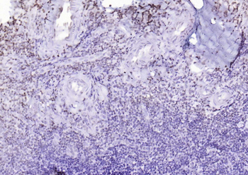
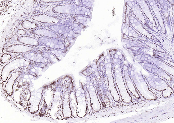
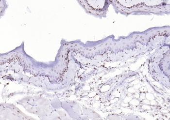
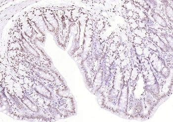
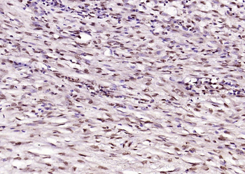
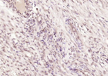
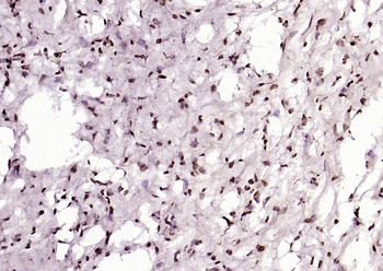
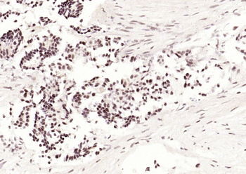
Images & Validation
−Item 1 of 3
| Tested Applications | ChIP, ELISA, IF, IHC, IP, Multiplex Assay, WB |
|---|---|
| Dilution Range | ELISA: 1:2,000 - 1:10,000, ChIP: 1 µg/µL at 4° o/n, IHC: 1:50, IF: 1:100-1:500, IP: 1:100, WB: 1:500 - 1:2,000 |
| Reactivity | Human |
| Application Notes |
Key Properties
−| Antibody Type | Primary Antibody |
|---|---|
| Host | Mouse |
| Clonality | Monoclonal |
| Isotype | IgG2a |
| Clone No. | BP53-12 |
| Immunogen | This protein A purified monoclonal antibody was produced by repeated immunizations with recombinant human p53 protein. |
| Target | TP53 |
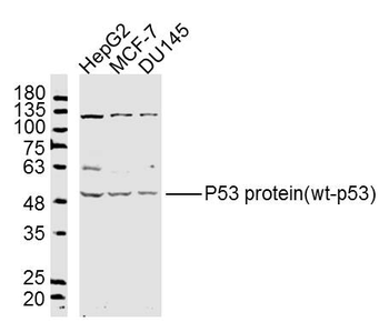
| Purity | This protein A purified mouse monoclonal antibody reacts specifically with p53 in human tissues and cell lines. The antibody recognizes a 53 kDa band corresponding to p53. Cross reactivity with p53 from other sources has not been determined. |
| Conjugation | Unconjugated |
Storage & Handling
−| Storage | Store vial at -20° C prior to opening. Aliquot contents and freeze at -20° C or below for extended storage. Avoid cycles of freezing and thawing. Centrifuge product if not completely clear after standing at room temperature. This product is stable for several weeks at 4° C as an undiluted liquid. Dilute only prior to immediate use. |
|---|---|
| Form/Appearance | Liquid (sterile filtered) |
| Buffer/Preservatives | Preservative: 0.01% (w/v) Sodium Azide. Stabilizer: None; Buffer: 0.02 M Potassium Phosphate, 0.5 M Sodium Chloride, pH 7.2 |
| Concentration | 1.0 mg/mL |
| Expiration Date | 12 months from date of receipt. |
| Dry Ice Shipping | Please note: This product requires shipment on dry ice. A dry ice surcharge will apply. |
| Disclaimer | For research use only |
Alternative Names
−mouse anti-p53 antibody, mouse anti-Tumor Suppressor p53 antibody, Phosphoprotein p53 antibody, TP53 antibody, Transformation related protein 53 antibody, TRP53 antibody, cellular Tumor antigen p53 antibody
Similar Products
−- Item 1 of 18
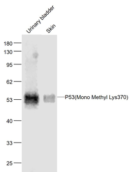
P53 (Mono Methyl Lys370) Mouse Monoclonal Antibody [orb500712]
IF, IHC-Fr, IHC-P, WB
Mouse, Rat
Human, Mouse, Rat
Mouse
Monoclonal
Unconjugated
50 μl, 100 μl, 200 μl, 200 μg - Item 1 of 12
P53 protein (wt-p53) Rabbit Polyclonal Antibody [orb11210]
FC, ICC, IF, IHC-Fr, IHC-P, WB
Bovine, Equine, Porcine, Rabbit, Sheep
Human, Mouse, Rat
Rabbit
Polyclonal
Unconjugated
- Item 1 of 11
P53 (Acetyl K382) Rabbit Polyclonal Antibody [orb11497]
FC, IF, IHC-Fr, IHC-P
Canine
Human
Rabbit
Polyclonal
Unconjugated
50 μl, 100 μl, 200 μl - Item 1 of 11
P53 (FL-393) Rabbit Polyclonal Antibody [orb2664]
FC, IF, IHC-Fr, IHC-P, WB
Bovine, Canine, Equine, Porcine, Sheep
Human, Monkey, Mouse, Rat
Rabbit
Polyclonal
Unconjugated
50 μl, 100 μl, 200 μl - Item 1 of 13
Phospho-P53 (Ser33) Rabbit Polyclonal Antibody [orb6596]
FC, ICC, IF, IHC-Fr, IHC-P, WB
Equine
Human, Mouse, Rat
Rabbit
Polyclonal
Unconjugated
50 μl, 100 μl, 200 μl


Quality Guarantee
Explore bioreagents carefree to elevate your research. All our products are rigorously tested for performance. If a product does not perform as described on its datasheet, our scientific support team will provide expert troubleshooting, a prompt replacement, or a refund. For full details, please see our Terms & Conditions and Buying Guide. Contact us at support@biorbyt.com.
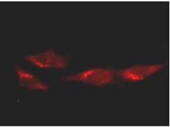
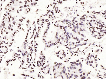
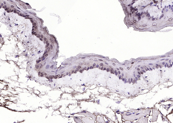
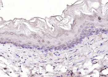
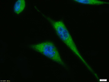
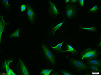
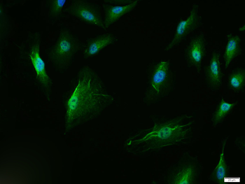
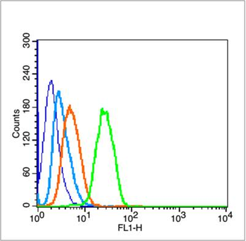
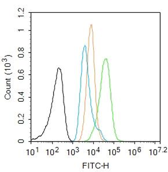
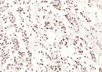
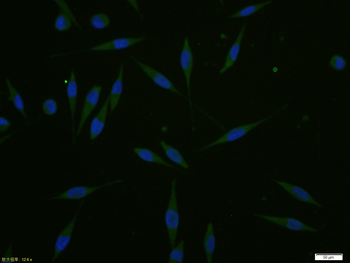
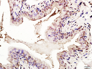
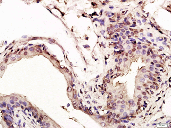
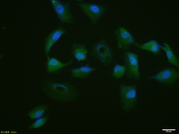
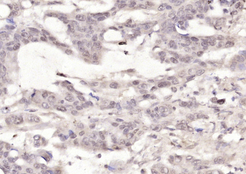
Immunofluorescence microscopy of HeLa cells using anti-p53. Biorbyt's Protein A purified Mab anti-p53 was used at a 1:100 dilution in 10% normal goat serum in PBS and reacted overnight at 4°C. After washes cells were incubated with a 1:500 dilution of AlexaFluor594 Goat-a-Mouse IgG diluted in normal goat serum for 1 h at room temperature.
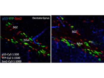
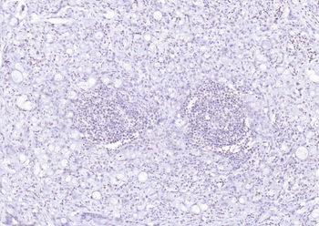
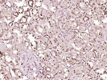
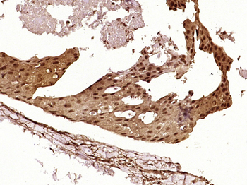
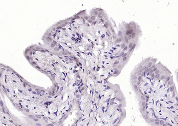
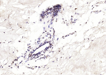
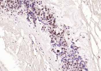
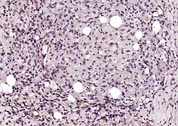
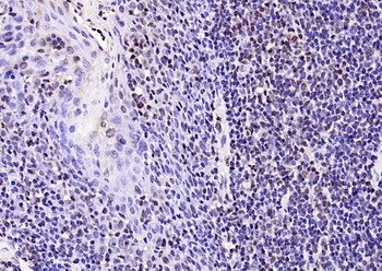
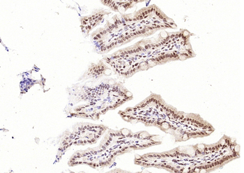
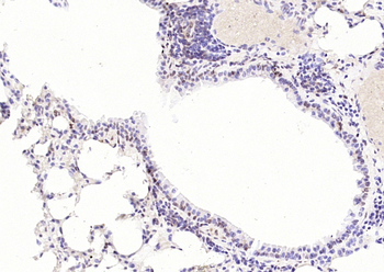
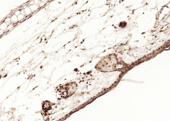
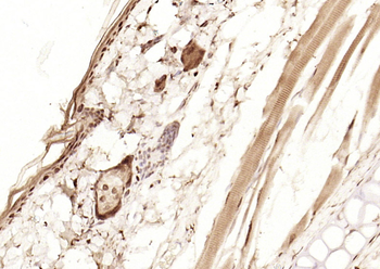
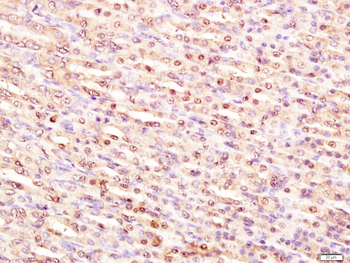
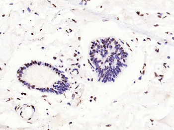
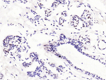
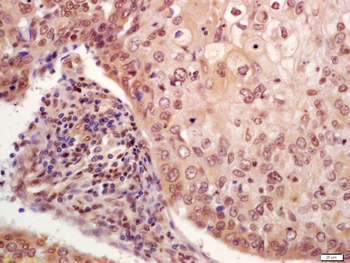
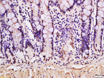
Immunofluorescence of Mouse anti-p53 antibody. Tissue: human brain. Fixation: free-floating. Antigen retrieval: not required. Primary antibody: anti-human-p53 antibody at 1:500 for 1 h at RT. Co-stained with YFP and Sox2 antibodies. Localization: p53 is nuclear and cytoplasmic. Staining: p53 as precipitated blue with Cy5, YFP as precipitated green with Cy2, and Sox2 as precipitated red with Cy3. z‐stacks from confocal expressed as one composite focal plain.
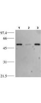
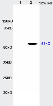
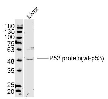
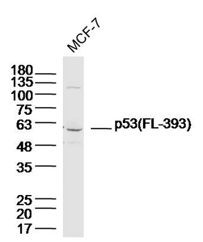
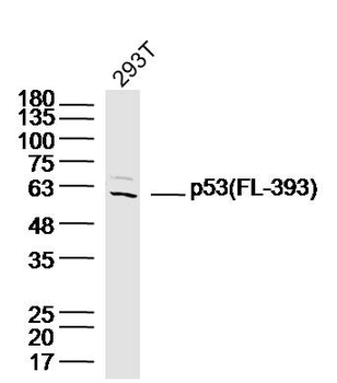
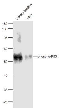
Western blotting using anti-p53 Antibody. Lane 1: HeLa whole cell lysate (p/n orb348668), Lane 2: HeLa cytosol fraction, and Lane 3: HeLa nuclear extract (p/n orb348671). Load of 15 µg was separated by 10% SDS-PAGE and transferred to nitrocellulose membrane. The membrane was blocked with 3% milk/TBST for 1 h at room temperature followed by incubation with Biorbyt's Protein A purified Mab anti-p53 antibody overnight at 4°C diluted 1:1500 in blocking solution. The membrane was washed 3X with TBST and then incubated with a 1:2000 dilution of HRP Goat-a-Mouse IgG (p/n orb347385) diluted in blocking buffer for 1 h at room temperature. After final washes the proteins reactive on the membrane were detected using ECL. Other detection systems will yield similar results.
Documents Download
Datasheet
Product Information
Request a Document
Protocol Information
WB
Western Blot (IB, immunoblot)
IHC
Immunohistochemistry
IF
Immunofluorescence
ELISA
Enzyme-linked Immunosorbent Assay (EIA)
IP
Immunoprecipitation
ChIP
Chromatin Immunoprecipitation
TP53 Antibody (orb344399)

- 0.0
Based on 0 reviews
Participating in our Biorbyt product reviews program enables you to support fellow scientists by sharing your firsthand experience with our products.
Login to Submit a ReviewAvailable Sizes
Select a size below
Choose Conjugation or Carrier Free Version
Free Secondary Antibody (20 ul)0/0
Please add an antibody product to your cart first.