You have no items in your shopping cart.
PDL2 Detection Set (Risk Free)
SKU: orb1227444
Description
Images & Validation
−Item 1 of 8
| Tested Applications | ELISA, FC, ICC, IF, IHC-P, WB |
|---|
Key Properties
−| Reactivity | Human |
|---|---|
| Assay Type | Detection |
| Concentration | Antibody 1 mg/mL |
| Conjugation | Unconjugated |
Storage & Handling
−| Storage | Antibodies can be stored at 4°C for three months and -20°C, stable for up to one year. As with all antibodies care should be taken to avoid repeated freeze thaw cycles. Antibodies should not be exposed to prolonged high temperatures. |
|---|---|
| Expiration Date | 6 months from date of receipt. |
| Disclaimer | For research use only |
Quality Guarantee
Explore bioreagents carefree to elevate your research. All our products are rigorously tested for performance. If a product does not perform as described on its datasheet, our scientific support team will provide expert troubleshooting, a prompt replacement, or a refund. For full details, please see our Terms & Conditions and Buying Guide. Contact us at support@biorbyt.com.
Western blot analysis of PD-L2 in overexpressing 293 cells using orb1239853, orb1239856, orb1239859, orb1238786, and orb1239860 antibody at 0.5 and 1 μg/ml, respectively. Larger molecular weight bands represent more highly post-translationally modified PD-L2.
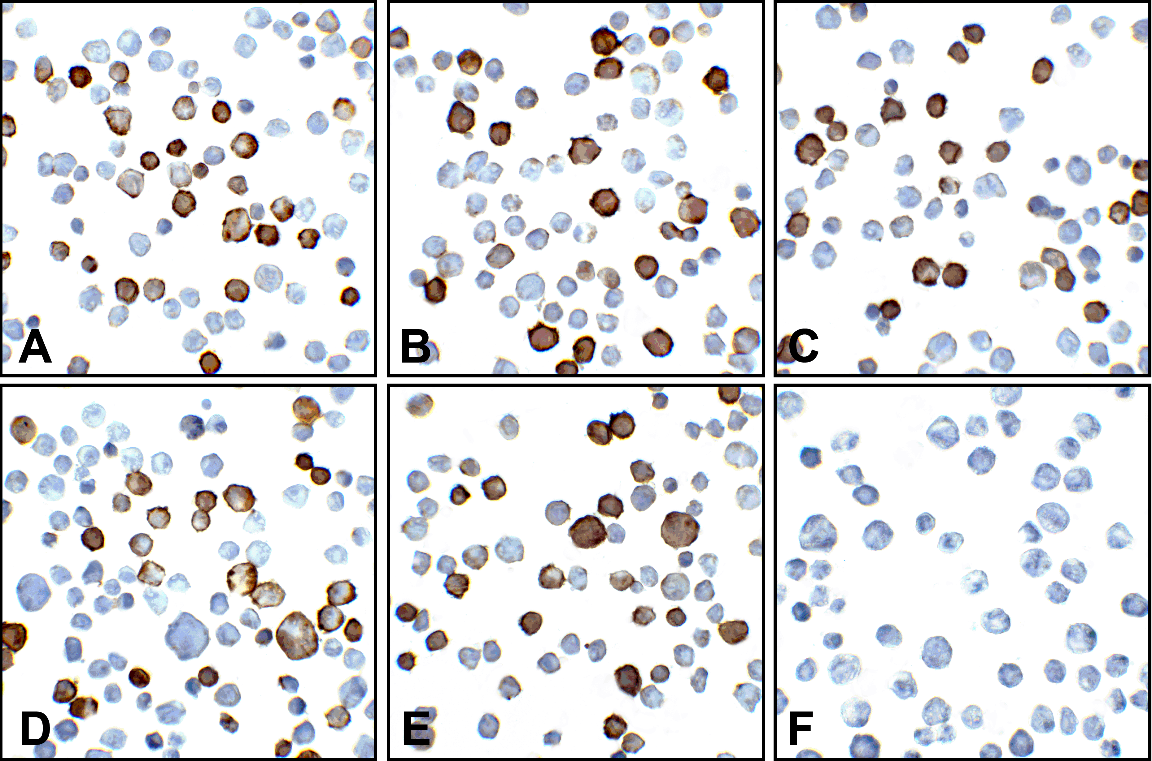
Immunocytochemistry of PD-L2 in overexpressing 293 cells using (A) orb1239853, (B) orb1239856, (C) orb1239859, (D) orb1238786, (E) orb1239860, and (F) control mouse IgG antibody at 5 μg/ml.
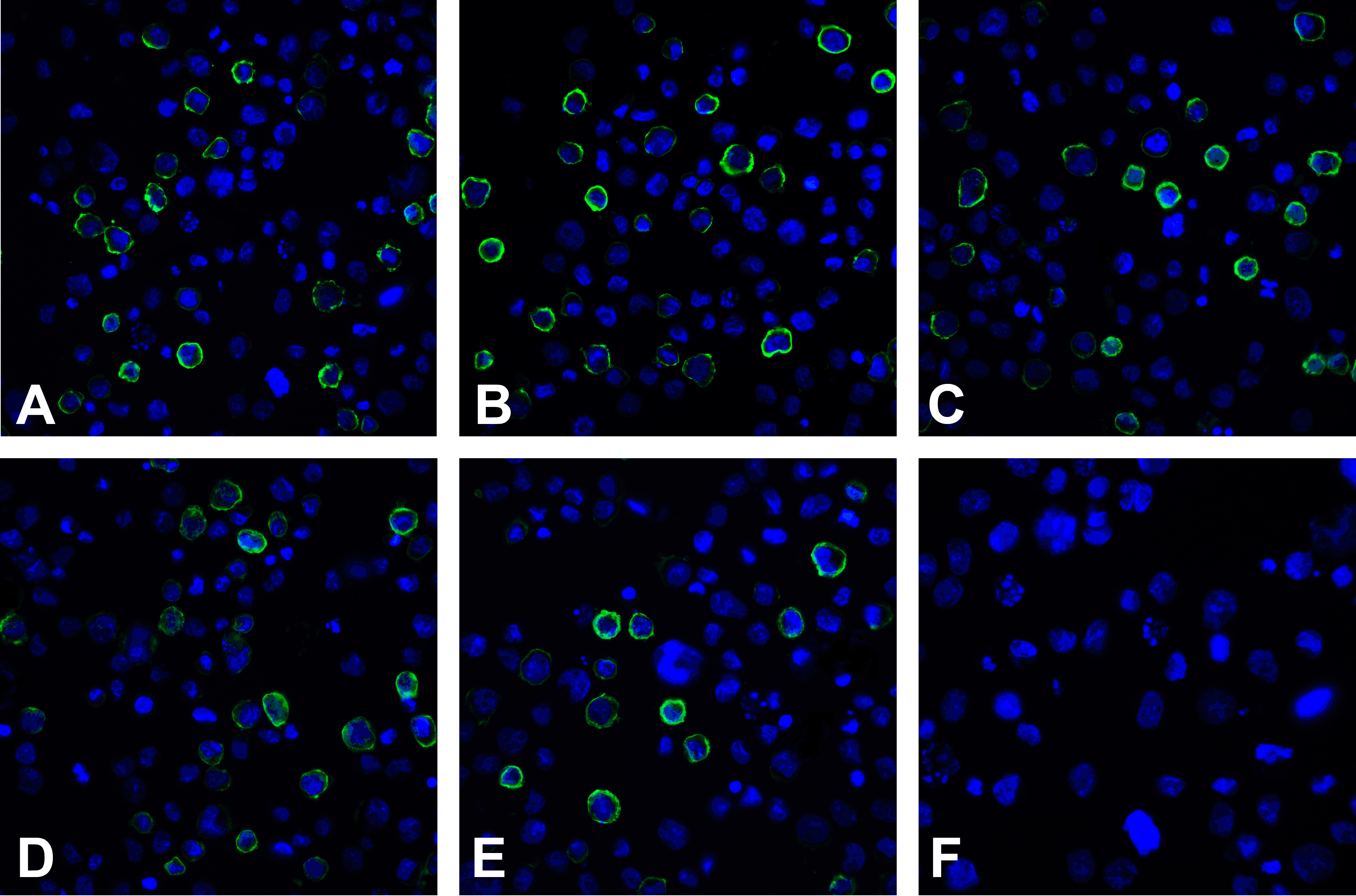
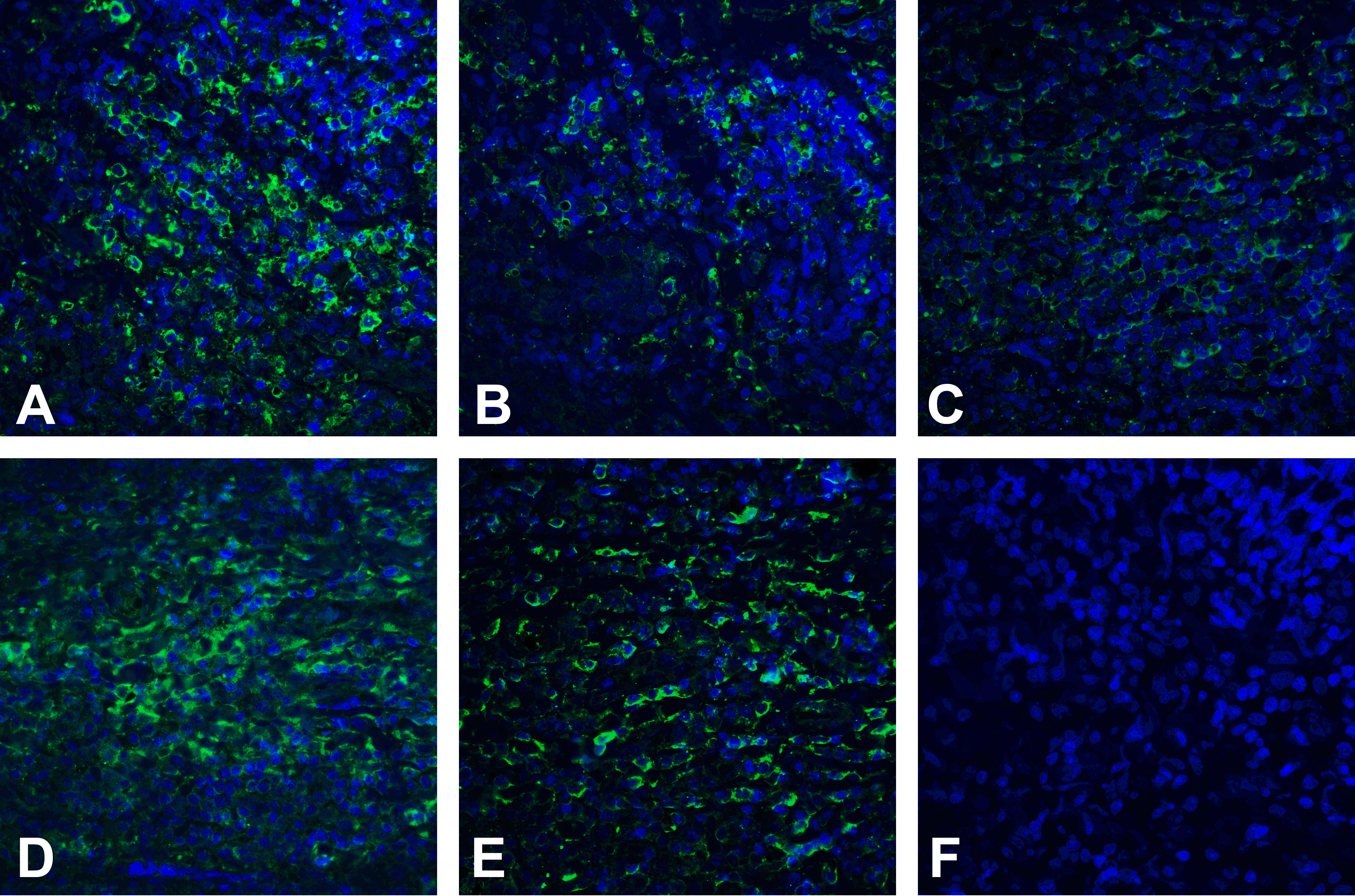
Immunofluorescence of PD-L2 in overexpressing 293 cells using (A) orb1239853, (B) orb1239856, (C) orb1239859, (D) orb1238786, (E) orb1239860, and (F) control mouse IgG antibody at 20 μg/ml.
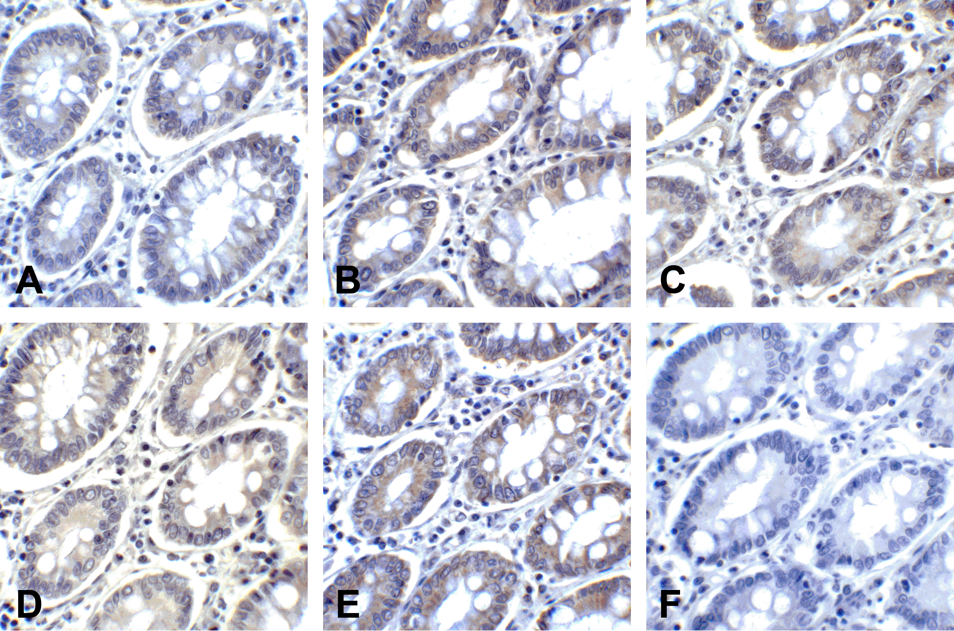
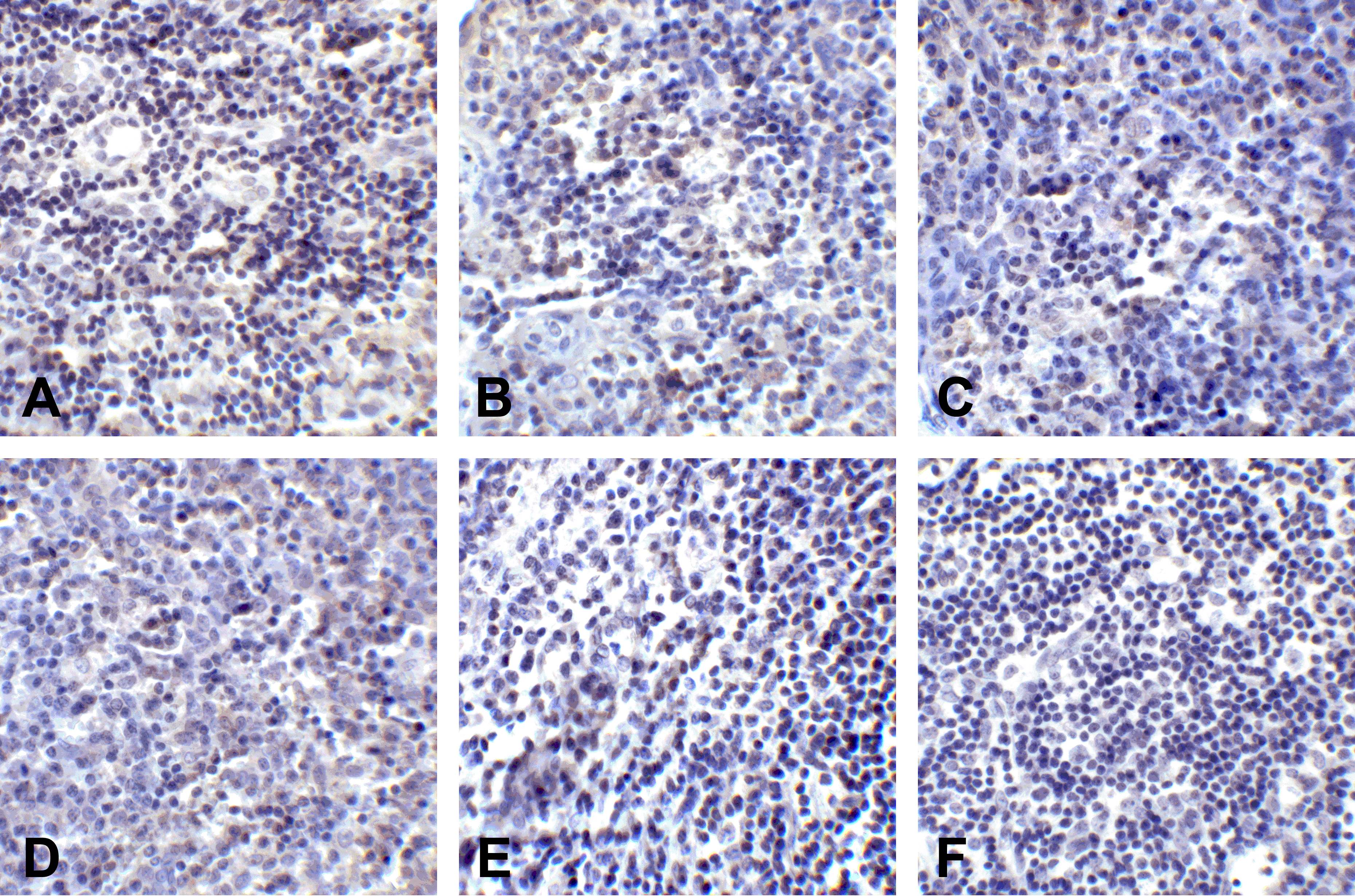
Immunohistochemistry of PD-L2 in human colon carcinoma tissue using (A) orb1239853, (B) orb1239856, (C) orb1239859, (D) orb1238786, (E) orb1239860, and (F) control mouse IgG antibody at 2 μg/ml.
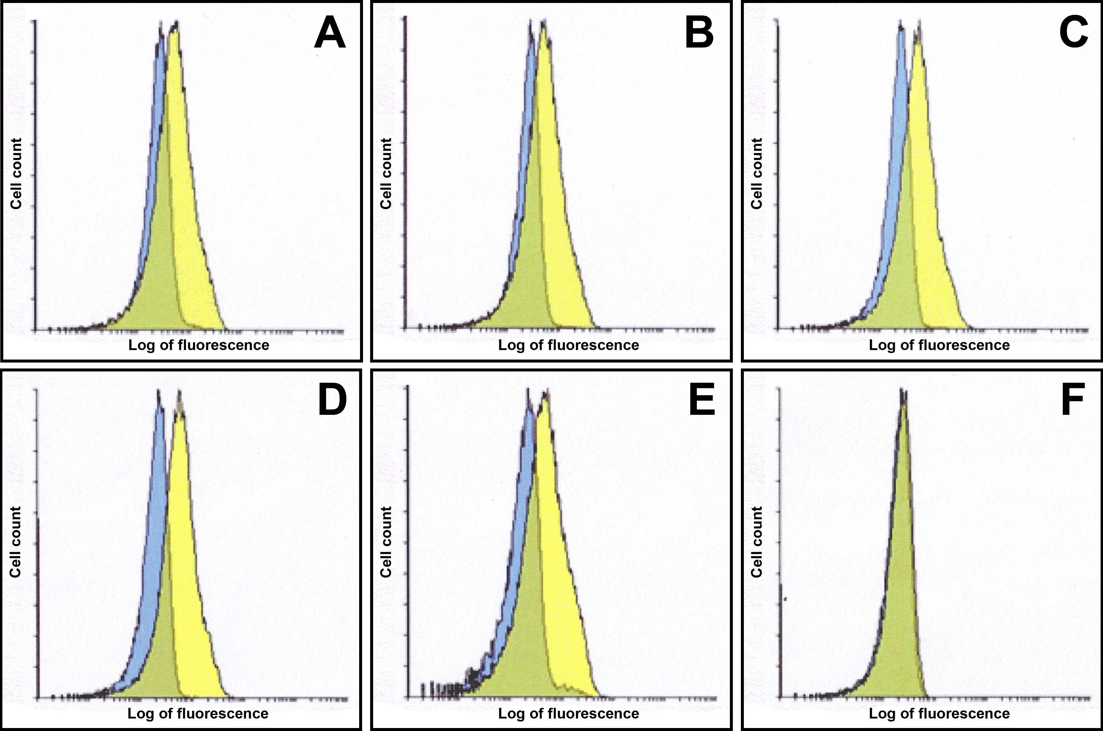
Flow cytometry analysis of PD-L2 overexpressing HEK293 cells using (A) orb1239853, (B) orb1239856, (C) orb1239859, (D) orb1238786, (E) orb1239860, and (F) control mouse IgG antibody at 10 μg/ml. Blue: Untransfected HEK293 cells. Yellow: PD-L2 overexpressing HEK293 cells.
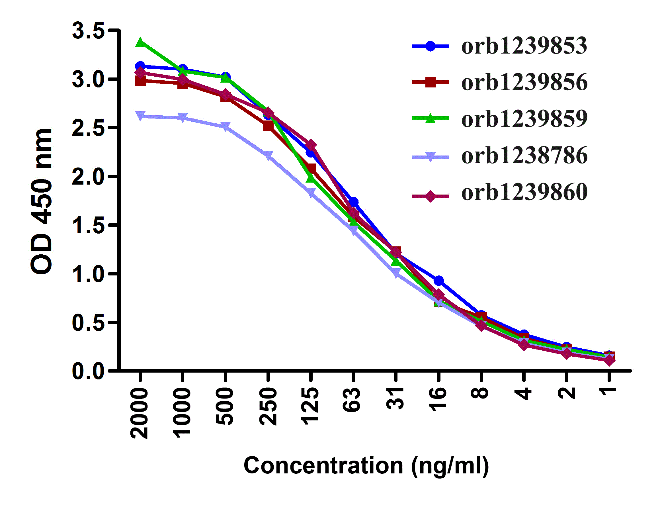
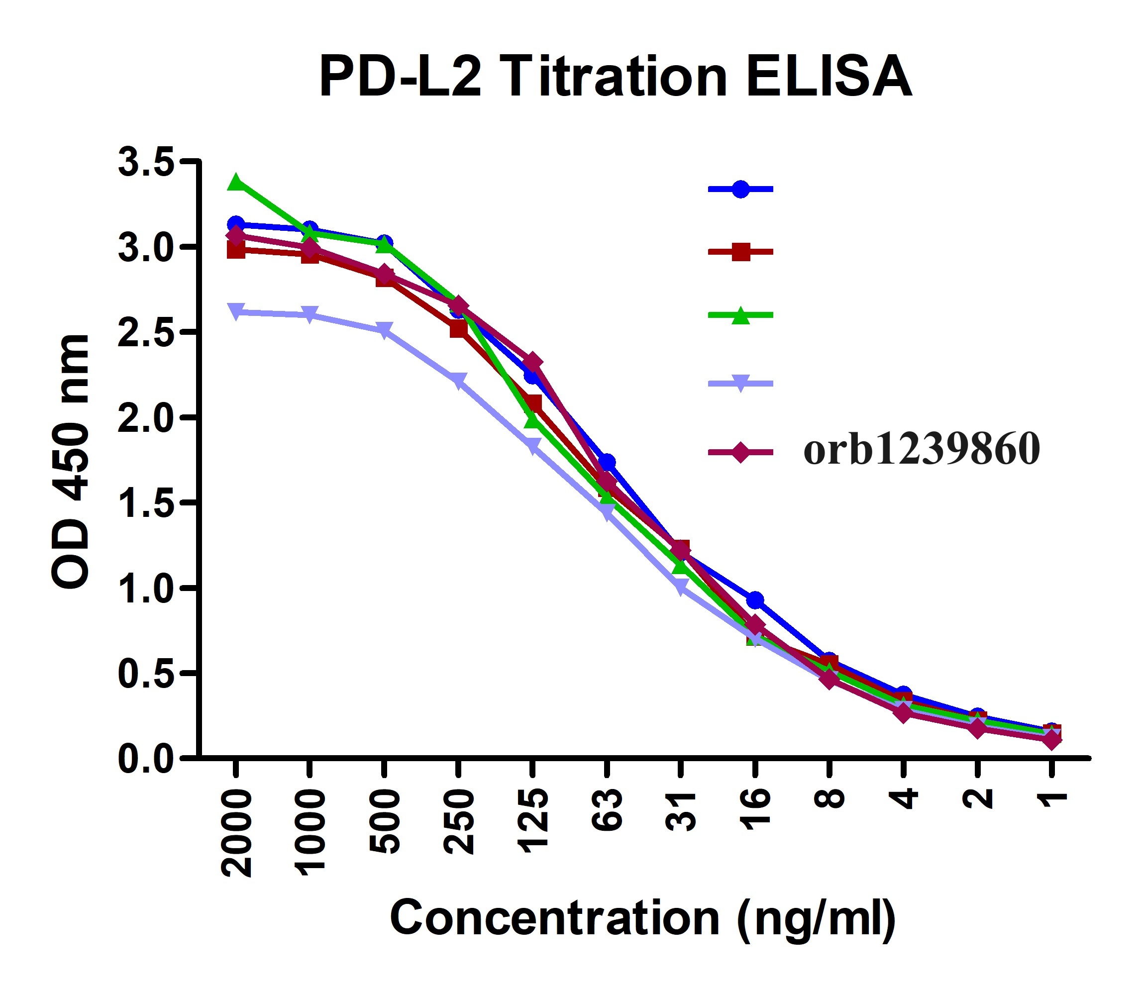
Titration curve analysis of PD-L2 mAbs to detect recombinant PD-L2 in ELISA with orb1239853, orb1239856, orb1239859, orb1238786, and orb1239860 abs at decreasing concentrations.
Immunofluorescence of PD-L2 in human tonsil tissue using (A) orb1239853, (B) orb1239856, (C) orb1239859, (D) orb1238786, (E) orb1239860, and (F) control mouse IgG antibody at 20 μg/ml.
Immunohistochemistry of PD-L2 in human tonsil tissue using (A) orb1239853, (B) orb1239856, (C) orb1239859, (D) orb1238786, (E) orb1239860, and (F) control mouse IgG antibody at 2 μg/ml.
Documents Download
Datasheet
Product Information
Request a Document
Protocol Information
WB
Western Blot (IB, immunoblot)
IHC-P
Immunohistochemistry Paraffin
FC
Flow Cytometry
IF
Immunofluorescence
ICC
Immunocytochemistry
ELISA
Enzyme-linked Immunosorbent Assay (EIA)
PDL2 Detection Set (Risk Free) (orb1227444)

- 0.0
Based on 0 reviews
Participating in our Biorbyt product reviews program enables you to support fellow scientists by sharing your firsthand experience with our products.
Login to Submit a ReviewAvailable Sizes
Select a size below