You have no items in your shopping cart.
Description
Research Area
Cancer Biology
Images & Validation
−Item 1 of 4
| Tested Applications | IF, IHC-P, WB |
|---|---|
| Dilution Range | IF - 1:200, WB - 1:500 |
| Reactivity | Human, Mouse |
Key Properties
−| Antibody Type | Primary Antibody |
|---|---|
| Host | Rabbit |
| Clonality | Polyclonal |
| Isotype | Rabbit IgG |
| Immunogen | This STAT3 Antibody is generated from rabbits immunized with a KLH conjugated synthetic phosphopeptide corresponding to amino acid residues surrounding S727 of human STAT3. |
| Target | STAT3 {ECO:0000303|PubMed:9630560, ECO:0000312|HGNC:HGNC:11364} |
| Molecular Weight | 88068 Da |
| Conjugation | Unconjugated |
Storage & Handling
−| Storage | Maintain refrigerated at 2-8°C for up to 2 weeks. For long term storage store at -20°C in small aliquots to prevent freeze-thaw cycles |
|---|---|
| Form/Appearance | Purified polyclonal antibody supplied in PBS with 0.09% (W/V) sodium azide. This antibody is purified through a protein A column, followed by peptide affinity purification. |
| Expiration Date | 12 months from date of receipt. |
| Disclaimer | For research use only |
Alternative Names
−Signal transducer and activator of transcription 3, Acute-phase response factor, STAT3, APRF
Similar Products
−- Item 1 of 1
Phospho-STAT3/STAT3 Rabbit Monoclonal Antibody [orb548555]
ICC, IF, IHC, IP, WB
Human, Mouse, Rat
Rabbit
Monoclonal
Unconjugated
100 μl 
Phospho-STAT3-S727 polyclonal antibody [orb1717483]
IHC, IP, WB
Human, Mouse, Rat
Rabbit
Polyclonal
Unconjugated
50 μl, 100 μlRabbit Phospho STAT3 (S727) Recombinant Monoclonal Antibody [orb1519593]
IP, WB
Human
Rabbit
Recombinant
Unconjugated
100 μg (BSA-free)
Quality Guarantee
Explore bioreagents carefree to elevate your research. All our products are rigorously tested for performance. If a product does not perform as described on its datasheet, our scientific support team will provide expert troubleshooting, a prompt replacement, or a refund. For full details, please see our Terms & Conditions and Buying Guide. Contact us at support@biorbyt.com.
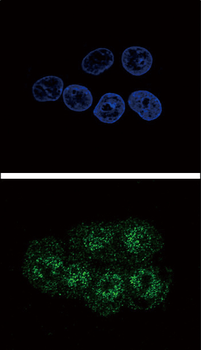
Confocal immunofluorescent analysis of Phospho-STAT3-S727 Antibody with HepG2 cell followed by Alexa Fluor 488-conjugated goat anti-rabbit lgG (green). DAPI was used to stain the cell nuclear (blue).
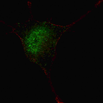
Fluorescent confocal image of SY5Y cells stained with phospho-STAT3-S727 antibody. SY5Y cells were fixed with 4% PFA (20 min), permeabilized with Triton X-100 (0.2%, 30 min). Cells were then incubated with phospho-STAT3-S727 primary antibody (1:100, 2 h at room temperature). For secondary antibody, Alexa Fluor 488 conjugated donkey anti-rabbit antibody (green) was used (1:1000, 1 h). Nuclei were counterstained with Hoechst 33342 (blue) (10 μg/ml, 5 min).
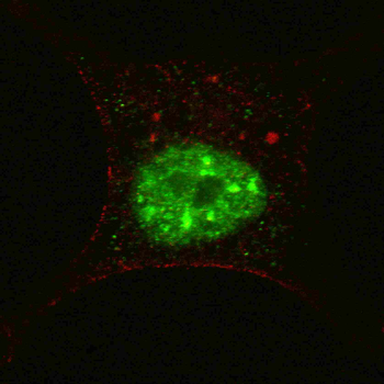
Fluorescent confocal image of SY5Y cells stained with phospho-STAT3-S727 antibody. SY5Y cells were fixed with 4% PFA (20 min), permeabilized with Triton X-100 (0.2%, 30 min). Cells were then incubated with phospho-STAT3-S727 primary antibody (1:200, 2 h at room temperature). For secondary antibody, Alexa Fluor 488 conjugated donkey anti-rabbit antibody (green) was used (1:1000, 1 h). Nuclei were counterstained with Hoechst 33342 (blue) (10 μg/ml, 5 min).
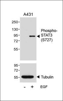
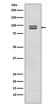
Western blot analysis of lysates from A431 cell line, untreated or treated with EGF, 100ng/ml, 5min, using Phospho-STAT3 (S727) Antibody (upper) or tubulin (lower).
Quick Database Links
Gene Symbol
STAT3 {ECO:0000303|PubMed:9630560, ECO:0000312|HGNC:HGNC:11364}
UniProt
RefSeq (Protein):NP_003141.2, NP_998827.1, NP_644805.1
UniProt Details
− No UniProt data available
NCBI Reference Sequences
−Associated Accession Numbers
Curated reference sequences for the gene transcript and protein product| Protein | NP_003141.2, NP_998827.1, NP_644805.1 |
|---|
Documents Download
Datasheet
Product Information
Request a Document
Protocol Information
WB
Western Blot (IB, immunoblot)
IHC-P
Immunohistochemistry Paraffin
IF
Immunofluorescence
Phospho-STAT3(S727) Antibody (orb1931219)

- 0.0
Based on 0 reviews
Participating in our Biorbyt product reviews program enables you to support fellow scientists by sharing your firsthand experience with our products.
Login to Submit a ReviewAvailable Sizes
Select a size below
Choose Conjugation or Carrier Free Version
Free Secondary Antibody (20 ul)0/0
Please add an antibody product to your cart first.