You have no items in your shopping cart.
Description
Research Area
Neuroscience
Images & Validation
−Item 1 of 3
| Tested Applications | IP, WB |
|---|---|
| Dilution Range | WB - 1:1,000 - 1:5,000; IP - 2 - 10 µg/mg lysate |
| Reactivity | Human, Mouse |
| Application Notes |
Key Properties
−| Antibody Type | Primary Antibody |
|---|---|
| Host | Rabbit |
| Clonality | Polyclonal |
| Isotype | IgG |
| Immunogen | Between 145 and C-terminus |
| Target | EXOSC1 |
| Purification | Antigen Affinity Purified |
| Conjugation | Unconjugated |
Storage & Handling
−| Storage | 2 - 8°C |
|---|---|
| Form/Appearance | Liquid |
| Buffer/Preservatives | Tris-citrate/phosphate buffer, pH 7 to 8 containing 0.09% Sodium Azide |
| Concentration | 1000 µg/ml |
| Expiration Date | 12 months from date of receipt. |
| Disclaimer | For research use only |
Alternative Names
−3'-5' exoribonuclease CSL4 homolog; CGI-108; CSL4; Csl4p; exosomal core protein CSL4; exosome complex component CSL4; Exosome component 1; homolog of yeast exosomal core protein CSL4; p13; SKI4; Ski4p
Similar Products
−- Item 1 of 1
- Item 1 of 2
EXOSC1 Rabbit Polyclonal Antibody [orb626850]
ELISA, IHC, WB
Human, Mouse, Rat
Rabbit
Polyclonal
Unconjugated
50 μg, 100 μg - Item 1 of 1
EXOSC1 Rabbit Polyclonal Antibody [orb411722]
WB
Human, Mouse
Rabbit
Polyclonal
Unconjugated
200 μl, 50 μl, 100 μl, 30 μl 
EXOSC1 Rabbit Polyclonal Antibody [orb1174788]
ELISA, WB
Mouse, Rat
Human
Rabbit
Polyclonal
Unconjugated
100 μgEXOSC1 Polyclonal Antibody [orb1419967]
ELISA, ICC, IF, IHC-Fr, IHC-P
Porcine, Rat
Rabbit
Polyclonal
Unconjugated
100 μl
Quality Guarantee
Explore bioreagents carefree to elevate your research. All our products are rigorously tested for performance. If a product does not perform as described on its datasheet, our scientific support team will provide expert troubleshooting, a prompt replacement, or a refund. For full details, please see our Terms & Conditions and Buying Guide. Contact us at support@biorbyt.com.

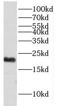
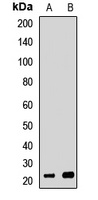
Detection of mouse EXOSC1 by western blot. Samples: Whole cell lysate (15 and 50 µg) from mouse NIH 3T3 cells. Antibodies: Affinity purified rabbit anti-EXOSC1 antibody orb1523997 used for WB at 1 µg/ml. Detection: Chemiluminescence with an exposure time of 3 minutes.

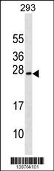
Detection of human EXOSC1 by western blot. Samples: Whole cell lysate from HeLa (15 and 50 µg) , HEK293T (50µg) , and Jurkat (50µg) cells. Antibodies: Affinity purified rabbit anti-EXOSC1 antibody orb1523997 used for WB at 0.4 µg/ml. Detection: Chemiluminescence with an exposure time of 3 minutes.

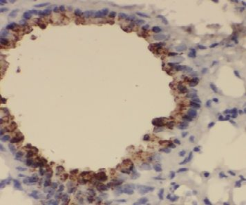
Detection of human EXOSC1 by western blot of immunoprecipitates. Samples: Whole cell lysate (1 mg for IP; 20% of IP loaded) from HeLa cells. Antibodies: Affinity purified rabbit anti-EXOSC1 antibody orb1523997 used for IP at 6 µg/mg lysate.
Quick Database Links
UniProt Details
− No UniProt data available
NCBI Reference Sequences
−Associated Accession Numbers
Curated reference sequences for the gene transcript and protein product| Protein | NP_057130.1 |
|---|
Documents Download
Datasheet
Product Information
Request a Document
Protocol Information
WB
Western Blot (IB, immunoblot)
IP
Immunoprecipitation
Rabbit EXOSC1 Antibody (orb1523997)

- 0.0
Based on 0 reviews
Participating in our Biorbyt product reviews program enables you to support fellow scientists by sharing your firsthand experience with our products.
Login to Submit a ReviewAvailable Sizes
Select a size below
Choose Conjugation or Carrier Free Version
Free Secondary Antibody (20 ul)0/0
Please add an antibody product to your cart first.