You have no items in your shopping cart.
Description
Research Area
Infectious Disease & Virology
Images & Validation
−Item 1 of 14
| Tested Applications | ELISA, IF, IHC, WB |
|---|---|
| Reactivity | Virus |
| Application Notes |
Key Properties
−| Antibody Type | Primary Antibody |
|---|---|
| Host | Rabbit |
| Clonality | Polyclonal |
| Isotype | IgG |
| Immunogen | Anti-SARS-CoV-2 (COVID-19) Spike antibody (orb1239976) was raised against a peptide corresponding to 20 amino acids near the carboxy terminus of SARS-CoV-2 (COVID-19) Spike glycoprotein. The immunogen is located within the last 50 amino acids of SARS-CoV-2 (COVID-19) Spike protein. |
| Target | S |
| Purification | SARS-CoV-2 (COVID-19) Spike Antibody is affinity chromatography purified via peptide column. |
| Conjugation | Unconjugated |
Storage & Handling
−| Storage | Maintain refrigerated at 2-8°C for up to 2 weeks. For long term storage store at -20°C in small aliquots to prevent freeze-thaw cycles. |
|---|---|
| Form/Appearance | Liquid |
| Buffer/Preservatives | SARS-CoV-2 (COVID-19) Spike Antibody is supplied in PBS containing 0.02% sodium azide. |
| Concentration | 1 mg/mL |
| Expiration Date | 12 months from date of receipt. |
| Disclaimer | For research use only |
Alternative Names
−SARS-CoV-2 (COVID-19) Spike Antibody: Severe acute respiratory syndrome coronavirus 2 (SARS-CoV-2), Surface Glycoprotein, Spike protein
Similar Products
−- Item 1 of 11
SARS-CoV-2 (COVID-19) Spike RBD Antibody [orb1239994]
ELISA, IF, IHC, WB
Virus
Rabbit
Polyclonal
Unconjugated
0.02 mg, 0.1 mg - Item 1 of 9
SARS-CoV-2 (COVID-19) Spike S1 Antibody [orb1239995]
ELISA, ICC, IF, IHC, WB
Virus
Rabbit
Polyclonal
Unconjugated
0.02 mg, 0.1 mg - Item 1 of 8
SARS-CoV-2 (COVID-19) Spike 681P Antibody [orb1239981]
ELISA, IF, WB
Virus
Rabbit
Polyclonal
Unconjugated
0.1 mg, 0.02 mg - Item 1 of 4
Anti-COVID-19 & SARS-CoV S glycoprotein [CR3022] [orb758974]
ELISA, IF
Virus
Human
Monoclonal
Unconjugated
0.05 mg - Item 1 of 4
Anti-COVID-19 & SARS-CoV S glycoprotein [CR3022] [orb758976]
ELISA, IF
Virus
Human
Monoclonal
Unconjugated
0.2 mg


![Anti-COVID-19 & SARS-CoV S glycoprotein [CR3022]](/images/pub/media/catalog/product/NewWebsite/35/orb758974_1.png)
![Anti-COVID-19 & SARS-CoV S glycoprotein [CR3022]](/images/pub/media/catalog/product/NewWebsite/35/orb758974_2.png)
![Anti-COVID-19 & SARS-CoV S glycoprotein [CR3022]](/images/pub/media/catalog/product/NewWebsite/35/orb758974_3.png)
![Anti-COVID-19 & SARS-CoV S glycoprotein [CR3022]](/images/pub/media/catalog/product/NewWebsite/35/orb758974_4.png)
![Anti-COVID-19 & SARS-CoV S glycoprotein [CR3022]](/images/pub/media/catalog/product/NewWebsite/35/orb758976_1.png)
![Anti-COVID-19 & SARS-CoV S glycoprotein [CR3022]](/images/pub/media/catalog/product/NewWebsite/35/orb758976_2.png)
![Anti-COVID-19 & SARS-CoV S glycoprotein [CR3022]](/images/pub/media/catalog/product/NewWebsite/35/orb758976_3.png)
![Anti-COVID-19 & SARS-CoV S glycoprotein [CR3022]](/images/pub/media/catalog/product/NewWebsite/35/orb758976_4.png)
Quality Guarantee
Explore bioreagents carefree to elevate your research. All our products are rigorously tested for performance. If a product does not perform as described on its datasheet, our scientific support team will provide expert troubleshooting, a prompt replacement, or a refund. For full details, please see our Terms & Conditions and Buying Guide. Contact us at support@biorbyt.com.
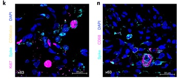
Immunofluorescent Validation of orb1239976 in SARS-CoV-2 Infected Lung Tissue (Singh et al., Nature Microbiology, 2021). Multilabel confocal immunofluorescence microscopy of formalin-fixed paraffin-embedded lung sections from rhesus macaques infected with SARS-CoV-2. SARS-CoV-2 spike-specific antibodies, orb1239976 (k, n) (turquoise) ; Ki67 (magenta) and neutrophil marker CD66abce (yellow) (k) ; pan-macrophage marker CD68 (magenta) (n) and DAPI (blue).
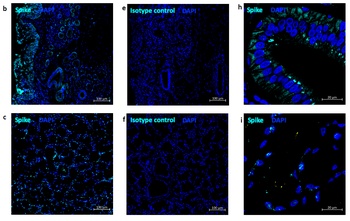
Immunofluorescent Validation of orb1239976 in SARS-CoV-2 Infected Nose and Tonsil (Singh et al., Nature Microbiology, 2021). Multi-label confocal immunofluorescence microscopy of nasal epithelium (20X-b, 63xh) and tonsil (20X-c, 63X-i) from rhesus macaques infected with SARS-CoV-2 with SARS-CoV-2 spike-specific antibodies, orb1239976 (turquoise), DAPI (blue). Rabbit IgG isotype control antibody wasused to stain the tissues to rule out any non-specific staining (e, f).
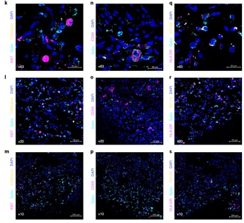
Immunofluorescent Validation of orb1239976 in SARS-CoV-2 Infected Lung Tissue (Singh et al., Nature Microbiology, 2021). Multilabel confocal immunofluorescence microscopy of formalin-fixed paraffin-embedded lung sections from rhesus macaques infected with SARS-CoV-2. SARS-CoV-2 spike-specific antibodies, orb1239976 (turquoise) ; Ki67 (magenta) and neutrophil marker CD66abce (yellow) (k-m) ; pan-macrophage marker CD68 (magenta) (n-p) ; HLA-DR (magenta) and pDC marker CD123 (yellow) (q–s) and DAPI (blue).

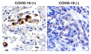
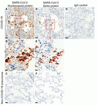
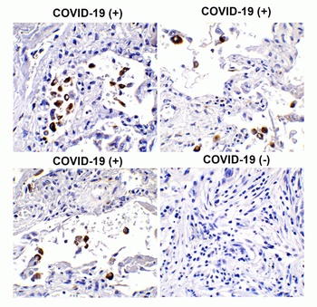
Immunohistochemistry Validation of SARS-CoV-2 (COVID-19) Spike in COVID-19 Patient Lung. Immunohistochemical analysis of paraffin-embedded COVID-19 patient lung tissue using anti-SARS-CoV-2 (COVID-19) Spike S2 antibody (orb1239976, 0.5 µg/mL). Tissue was fixed with formaldehyde and blocked with 10% serum for 1 h at RT; antigen retrieval was by heat mediation with a citrate buffer (pH6). Samples were incubated with primary antibody overnight at 4 °C. A goat anti-rabbit IgG H&L (HRP) at 1/250 was used as secondary. Counter stained with Hematoxylin. Strong spike protein signal was observed in macrophages and airway epithelium of COVID-19 patient lung, but not in non-COVID-19 patient lung.
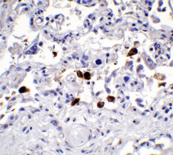
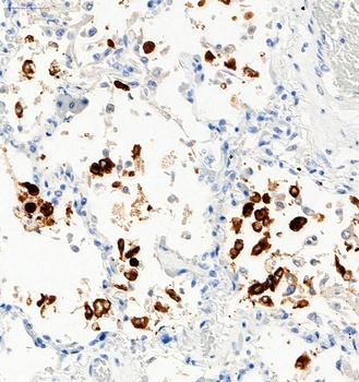
Immunohistochemistry Validation of SARS-CoV-2 (COVID-19) Spike in COVID-19 Patient Lung. Immunohistochemical analysis of paraffin-embedded COVID-19 patient lung tissue using anti-SARS-CoV-2 (COVID-19) Spike S2 antibody (orb1239976, 0.5 µg/mL). Tissue was fixed with formaldehyde and blocked with 10% serum for 1 h at RT; antigen retrieval was by heat mediation with a citrate buffer (pH6). Samples were incubated with primary antibody overnight at 4 °C. A goat anti-rabbit IgG H&L (HRP) at 1/250 was used as secondary. Counter stained with Hematoxylin.Strong spike protein signal was observed in macrophages of COVID-19 patient lung.
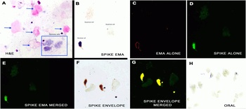
IHC/IF Validation in COVID-19 Patient Sample. (Nuovo et al., 2020) Detection of SARS-CoV-2 proteins in nasopharyngeal swab cell preparations. B. An intense signal for covid-19 spike protein tested by SARS-CoV-2 spike antibodies (orb1239976) was observed in the glandular cells. F-H. Co-expression of spike detected by spike antibodies (orb1239976, 0.2 µg/mL) and envelope proteins detected by envelope antibodies (orb1239971, 2 µg/mL) of SARS-CoV-2 (F) documented localization of each protein to glandular cells (G, yellow). No signal was seen in oral swabs of positive cases (H). Both the spike and envelope protein detected by anti-spike antibodies (orb1239976) and anti-envelope antibodies (orb1239971) produced a signal in the nasopharyngeal swabs of the three cases and no signal was evident in the nasopharyngeal swabs of the seven controls.
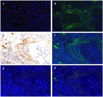
IF Validation of SARS-CoV2 Spike in COVID-19 Patient Lung. (Magro et al., 2020) SARS-CoV2 spike protein (red, panel C) detected detected by anti-spike antibodies (orb1239976, 0.2 µg/mL) colocalized with C4d (green in panel d, merged in yellow). Spike protein (red, panel g) was also colocalized with C5b-9 (green in panel f&h, merged in yellow).
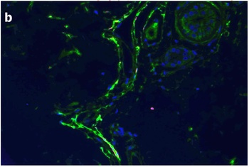
IF Validation of SARS-CoV2 Spike in COVID-19 Patient Skin. (Magro et al., 2020) C4d is highlighted green while COVID-19 spike protein detected by anti-spike antibodies (orb1239976, 0.2 µg/mL) shows a red staining pattern; a yellow signal is discernible indicative of co-localization of C4d and viral protein within the microvasculature.
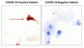
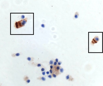
IHC Validation of SARS-CoV2 Spike in the Nasopharyngeal Swab Sample of the COVID-19 Patient. Strong spike signal was detected by anti-spike antibodies (orb1239976, 0.2 µg/mL) in the nasopharyngeal swab sample of the COVID-19 patient and no spike signal was observed in the sample of the COVID-19 negative patient. COVID-19 cases were confirmed by PCR.
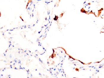
IHC Validation of SARS-CoV2 Spike in COVID-19 Patient Lung. Strong spike signal was detected by anti-spike antibodies (orb1239976, 0.2 µg/mL) in the lung of the COVID-19 patient confirmed by PCR.
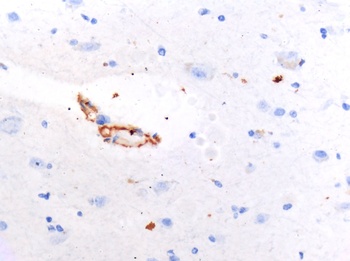
IHC Validation of SARS-CoV2 Spike in COVID-19 Patient Brain. Spike protein was detected by anti-spike antibodies (orb1239976, 0.2 µg/mL) in the brain of the COVID-19 patient confirmed by PCR.
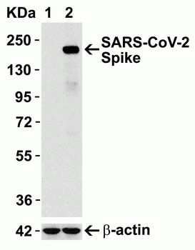
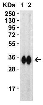
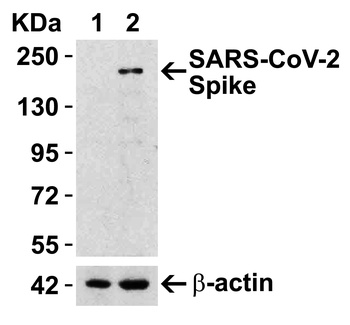
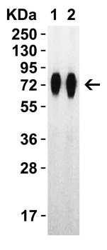
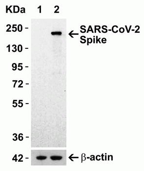
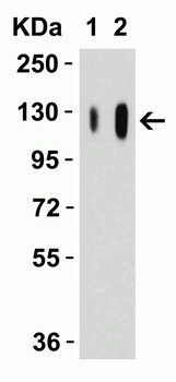
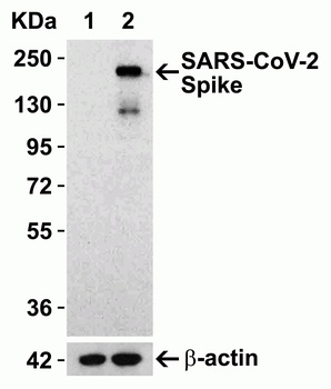
Overexpression Validation in Spike Transfected 293 Cells. Loading: 15 µg per lane of 293 cell lysate. Antibodies: SARS-CoV-2 (COVID-19) Spike, orb1239976 (1 µg/mL), 1h incubation at RT in 5% NFDM/TBST. Secondary: Goat anti-rabbit IgG HRP conjugate at 1:10000 dilution. Lane 1: WT 293 cells and Lane 2: SARS-CoV-2 Spike overexpressed 293 cells.
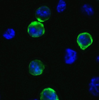
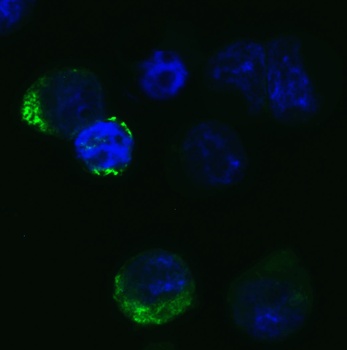
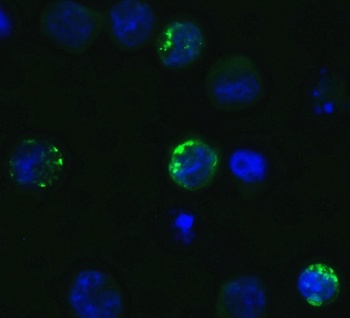
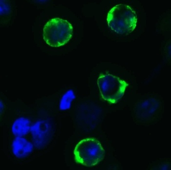
Immunofluorescence Validation of SARS-CoV-2 (COVID-19) Spike in 293T Cells. Immunofluorescent analysis of 4% paraformaldehyde-fixed 293T cells labeling SARS-CoV-2 (COVID-19) Spike with orb1239976 at 1 µg/mL, followed by goat anti-rabbit IgG secondary antibody at 1/500 dilution (green) and DAPI staining (blue).
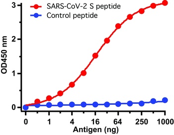
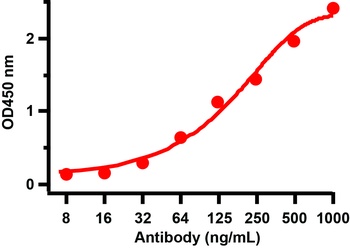
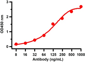
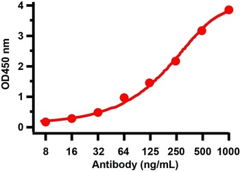
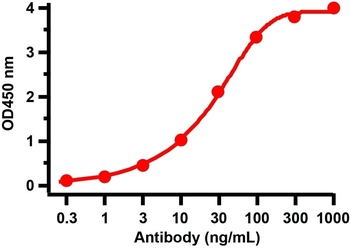
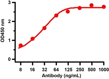
ELISA Test. Antibodies: SARS-CoV-2 (COVID-19) Spike antibody, orb1239976 (1 µg/mL). A direct ELISA was performed using immunogen or control peptide as coating antigen and the anti-SARS-CoV-2 (COVID-19) Spike antibody as the capture antibody. Secondary: Goat anti-rabbit IgG HRP conjugate at 1:20000 dilution. Detection range is from 0.5 ng/mL to 1000 ng/mL.
Documents Download
Datasheet
Product Information
Request a Document
Protocol Information
WB
Western Blot (IB, immunoblot)
IHC
Immunohistochemistry
IF
Immunofluorescence
ELISA
Enzyme-linked Immunosorbent Assay (EIA)
SARS-CoV-2 (COVID-19) Spike Antibody (orb1239976)

- 0.0
Based on 0 reviews
Participating in our Biorbyt product reviews program enables you to support fellow scientists by sharing your firsthand experience with our products.
Login to Submit a ReviewAvailable Sizes
Select a size below
Free Secondary Antibody (20 ul)0/0
Please add an antibody product to your cart first.