You have no items in your shopping cart.
SMAD3 phospho T179 Antibody
SKU: orb345687
Description
Images & Validation
−Item 1 of 2
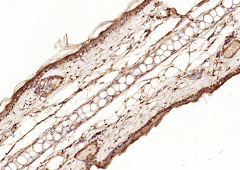
| Tested Applications | ELISA, WB |
|---|---|
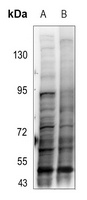
| Dilution Range | ELISA: 1:15,000-1:75,000, WB: 1:1,000 |
| Reactivity | Mouse |
| Application Notes |
Key Properties
−| Antibody Type | Primary Antibody |
|---|---|
| Host | Rabbit |
| Clonality | Polyclonal |
| Isotype | IgG |
| Immunogen | Anti-SMAD3 pT179 antibody was prepared by repeated immunizations with a synthetic peptide corresponding to an internal region of human Smad3 protein surrounding amino acid residue 179. |
| Purity | Anti-SMAD3 pT179 affinity-purified antibody is directed against the phosphorylated form of human Smad3 protein at the pT179 residue. The product was affinity purified from monospecific antiserum by immunoaffinity purification. Antiserum was first purified against the phosphorylated form of the immunizing peptide. The resultant affinity purified antibody was then cross adsorbed against the non-phosphorylated form of the immunizing peptide. Reactivity occurs against human Smad3 pT179 protein and the antibody is specific for the phosphorylated form of the protein. Reactivity with non-phosphorylated human Smad3 is minimal by ELISA and western blot. Expect reactivity against phosphorylated Smad2. Reactivity against other phosphorylated Smad family members is not known. A BLAST analysis was used to suggest cross reactivity with Smad3 from human, mouse, rat, pig, dog, and marmoset based on 100% sequence homology with the immunogen. Reactivity against homologues from other sources is not known. |
| Conjugation | Unconjugated |
Storage & Handling
−| Storage | Store vial at -20° C or below prior to opening. This vial contains a relatively low volume of reagent (25 µL). To minimize loss of volume dilute 1:10 by adding 225 µL of the buffer stated above directly to the vial. Recap, mix thoroughly and briefly centrifuge to collect the volume at the bottom of the vial. Use this intermediate dilution when calculating final dilutions as recommended below. Store the vial at -20°C or below after dilution. Avoid cycles of freezing and thawing. |
|---|---|
| Form/Appearance | Liquid (sterile filtered) |
| Buffer/Preservatives | Preservative: 0.01% (w/v) Sodium Azide. Stabilizer: None; Buffer: 0.02 M Potassium Phosphate, 0.15 M Sodium Chloride, pH 7.2 |
| Concentration | 1.1 mg/mL |
| Expiration Date | 12 months from date of receipt. |
| Dry Ice Shipping | Please note: This product requires shipment on dry ice. A dry ice surcharge will apply. |
| Disclaimer | For research use only |
Alternative Names
−rabbit anti-SMAD3 pT179 antibody, SMAD-3, SMAD 3, mothers against decapentaplegic homolog 3 antibody, MAD homolog 3, Mothers against DPP homolog 3, SMAD family member 3, MADH3, MADH 3, JV15-2
Similar Products
−- Item 1 of 13
Phospho-Smad3 (Thr179) Rabbit Polyclonal Antibody [orb313112]
FC, ICC, IF, IHC-Fr, IHC-P
Bovine, Canine, Equine, Porcine, Sheep
Human, Mouse, Rat
Rabbit
Polyclonal
Unconjugated
50 μl, 100 μl, 200 μl - Item 1 of 2
SMAD3 (Phospho-T179) Rabbit Polyclonal Antibody [orb304595]
IHC, WB
Canine, Human, Monkey, Mouse, Porcine, Rat, Sheep
Rabbit
Polyclonal
Unconjugated
30 μl, 100 μl, 200 μl, 50 μl - Item 1 of 2
Smad3 (phospho-T179) polyclonal antibody [orb642405]
WB
Human, Mouse, Rat
Rabbit
Polyclonal
Unconjugated
25 μl, 200 μl, 100 μl, 50 μlPhospho-Smad3 (Thr179) Rabbit Polyclonal Antibody (FITC) [orb502703]
FC, ICC, IF
Bovine, Canine, Equine, Porcine, Sheep
Human, Mouse, Rat
Rabbit
Polyclonal
FITC
100 μl


Quality Guarantee
Explore bioreagents carefree to elevate your research. All our products are rigorously tested for performance. If a product does not perform as described on its datasheet, our scientific support team will provide expert troubleshooting, a prompt replacement, or a refund. For full details, please see our Terms & Conditions and Buying Guide. Contact us at support@biorbyt.com.

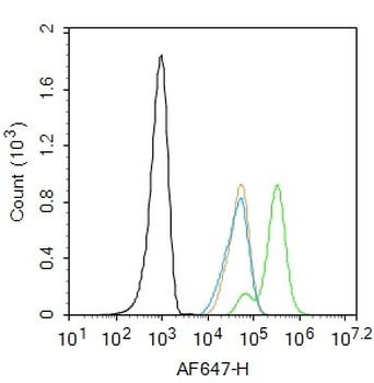
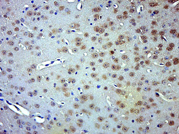
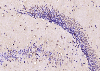
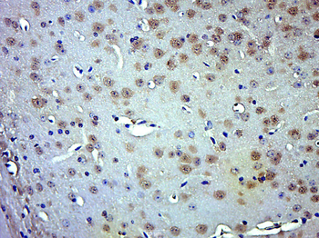
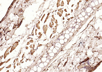
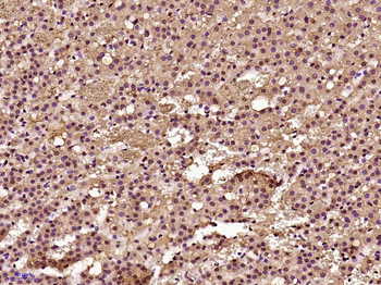
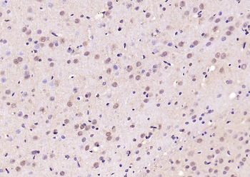
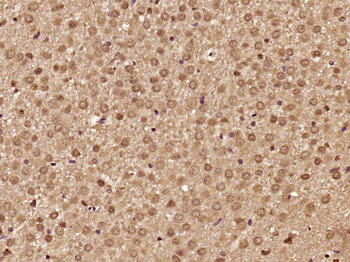
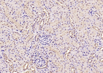
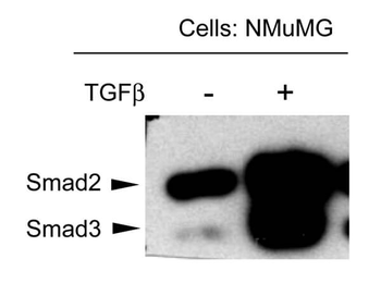
NMuMG mouse mammary epithelial cells were probed for the activation of Smad3 by detecting phosphorylation of threonine 179. The cells were either untreated or treated with TGF-beta, transferred to membranes and probed with Anti-SMAD3 pT179 (RABBIT) Antibody. The antibody detects only Smad3 in stimulated cells suggesting detection of phosphorylated SMAD3 at T179.
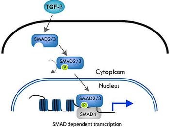
The SMAD pathway follows the canonical TGF-ß signaling pathway. TGF-ß dimers bind to a receptor thereby activating the pathway. The type I receptor then recruits and phosphorylates a receptor regulated SMAD (R-SMAD).i.e. SMAD2 or SMAD3. The R-SMAD then binds to the common SMAD (coSMAD) i.e. SMAD4, and forms a heterodimeric complex. This complex then enters the cell nucleus and acts as a transcription factor.
Documents Download
Datasheet
Product Information
Request a Document
Protocol Information
WB
Western Blot (IB, immunoblot)
ELISA
Enzyme-linked Immunosorbent Assay (EIA)
SMAD3 phospho T179 Antibody (orb345687)

- 0.0
Based on 0 reviews
Participating in our Biorbyt product reviews program enables you to support fellow scientists by sharing your firsthand experience with our products.
Login to Submit a Review