You have no items in your shopping cart.
ACTA1 Antibody
SKU: orb1939320
Description
Research Area
Cancer Biology, Cardiovascular Research, Immunology & Inflammation, Signal Transduction
Images & Validation
−Item 1 of 5
| Tested Applications | FC, IHC-P, WB |
|---|---|
| Dilution Range | WB - 1:1000 |
| Reactivity | Human |
| Predicted Reactivity | Bovine, Gallus, Mouse, Porcine, Rabbit, Rat |
Key Properties
−| Antibody Type | Primary Antibody |
|---|---|
| Host | Mouse |
| Clonality | Monoclonal |
| Isotype | IgG1,k |
| Molecular Weight | 42051 Da |
| Conjugation | Unconjugated |
Storage & Handling
−| Storage | Maintain refrigerated at 2-8°C for up to 2 weeks. For long term storage store at -20°C in small aliquots to prevent freeze-thaw cycles |
|---|---|
| Form/Appearance | Purified monoclonal antibody supplied in PBS with 0.09% (W/V) sodium azide. This antibody is purified through a protein G column, followed by dialysis against PBS. |
| Expiration Date | 12 months from date of receipt. |
| Disclaimer | For research use only |
Alternative Names
−ACTA
Similar Products
−- Item 1 of 11
Pan Actin Recombinant Rabbit Monoclonal Antibody [orb1172771]
FC, ICC, IF, IHC-Fr, IHC-P, WB
Human, Mouse, Rat
Human, Mouse, Rat
Rabbit
Recombinant
Unconjugated
25 μl, 50 μl, 100 μl - Item 1 of 6
Actin Rabbit Polyclonal Antibody [orb182438]
IF, IHC-Fr, IHC-P, WB
Bovine, Canine, Human, Rabbit
Mouse, Rat
Rabbit
Polyclonal
Unconjugated
200 μl, 50 μl, 100 μl - Item 1 of 7
ACTA2 Antibody [orb389300]
IHC, WB
Human, Rabbit, Rat
Mouse
Monoclonal
Unconjugated
100 μg, 100 μg (without BSA and Azide), 20 μg - Item 1 of 8
Actin/ACTA1 Rabbit Polyclonal Antibody [orb259587]
IHC, WB
Human, Mouse, Rat
Rabbit
Polyclonal
Unconjugated
100 μg - Item 1 of 5
ACTA2 Antibody [orb389301]
FC, IF, IHC
Canine, Feline, Human, Rabbit, Rat
Mouse
Monoclonal
Unconjugated
20 μg, 100 μg, 100 μg (without BSA and Azide)


Quality Guarantee
Explore bioreagents carefree to elevate your research. All our products are rigorously tested for performance. If a product does not perform as described on its datasheet, our scientific support team will provide expert troubleshooting, a prompt replacement, or a refund. For full details, please see our Terms & Conditions and Buying Guide. Contact us at support@biorbyt.com.
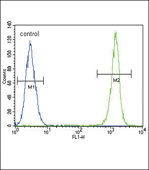
ACTA1 Antibody flow cytometric analysis of A549 cells (right histogram) compared to a negative control cell (left histogram). FITC-conjugated goat-anti-mouse secondary antibodies were used for the analysis.
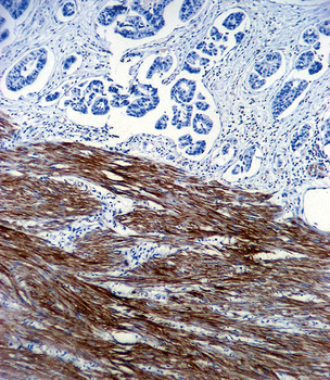
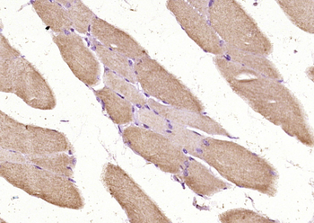
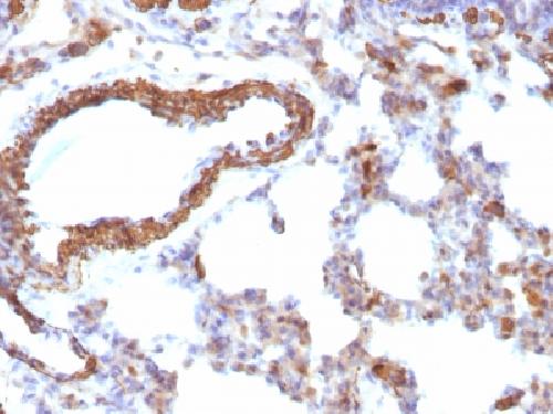
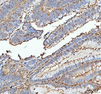
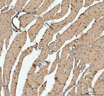
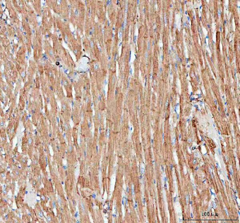
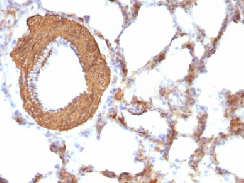
ACTA1 Antibody immunohistochemistry analysis in formalin fixed and paraffin embedded human colon carcinoma followed by peroxidase conjugation of the secondary antibody and DAB staining. This data demonstrates the use of ACTA1 Antibody for immunohistochemistry. Clinical relevance has not been evaluated.
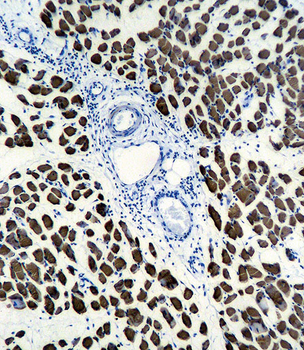
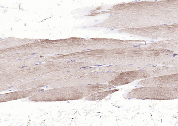
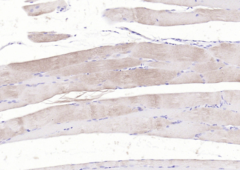
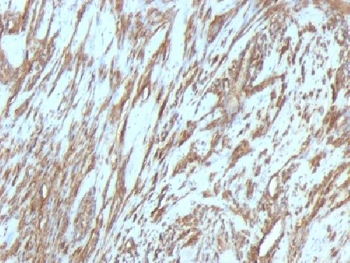
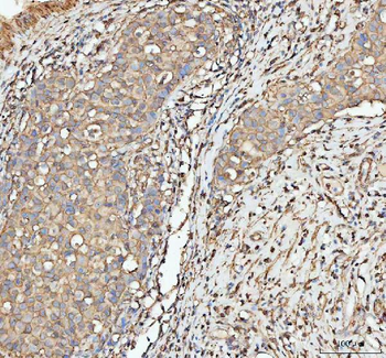
ACTA1 Antibody immunohistochemistry analysis in formalin fixed and paraffin embedded human skeletal muscle followed by peroxidase conjugation of the secondary antibody and DAB staining. This data demonstrates the use of ACTA1 Antibody for immunohistochemistry. Clinical relevance has not been evaluated.
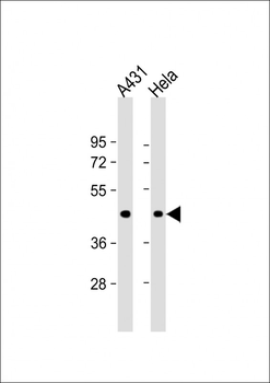
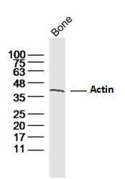
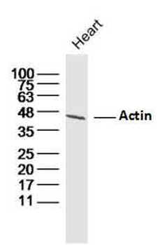
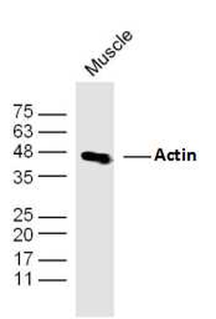
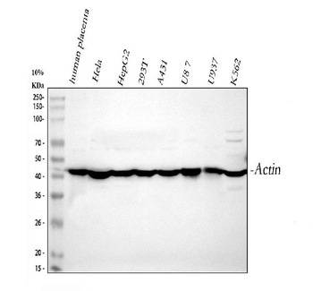
All lanes: Anti- at 1:1000 dilution. Lane 1: A431 whole cell lysate. Lane 2: Hela whole cell lysate.Lysates/proteins at 20 µg per lane. Secondary Goat Anti-mouse IgG, (H+L), Peroxidase conjugated at 1/10000 dilution. Predicted band size: 42 kDa. Blocking/Dilution buffer: 5% NFDM/TBST.
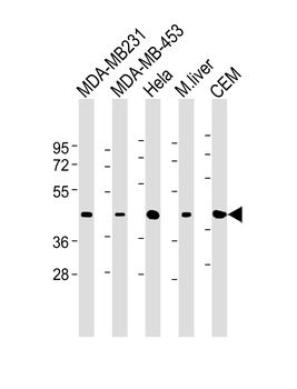
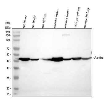
All lanes: Anti-ACTA1 Antibody at 1:500-1:1000 dilution. Lane 1: MDA-MB231 whole cell lysate. Lane 2: MDA-MB-453 whole cell lysate. Lane 3: Hela whole cell lysate. Lane 4: mouse liver lysate. Lane 5: CEM whole cell lysate. Secondary Goat Anti-mouse IgG, (H+L), Peroxidase conjugated at 1/10000 dilution. Predicted band size: 42051 Da. Blocking/Dilution buffer: 5% NFDM/TBST.
Quick Database Links
UniProt
UniProt Details
− No UniProt data available
Documents Download
Datasheet
Product Information
Request a Document
Protocol Information
WB
Western Blot (IB, immunoblot)
IHC-P
Immunohistochemistry Paraffin
FC
Flow Cytometry
ACTA1 Antibody (orb1939320)

- 0.0
Based on 0 reviews
Participating in our Biorbyt product reviews program enables you to support fellow scientists by sharing your firsthand experience with our products.
Login to Submit a ReviewAvailable Sizes
Select a size below