You have no items in your shopping cart.
Description
Research Area
Disease Biomarkers
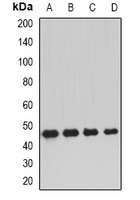
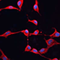
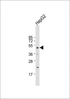
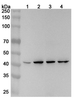
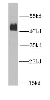
Images & Validation
−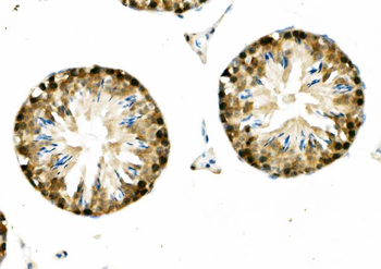

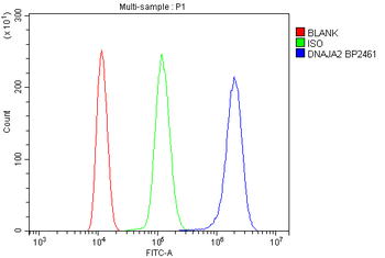
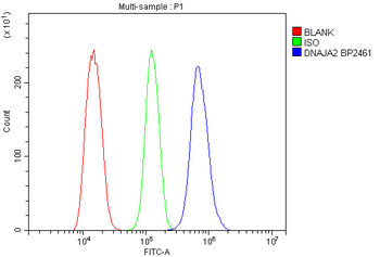
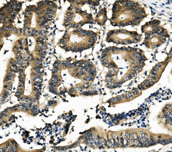
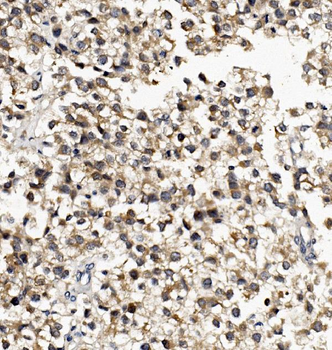
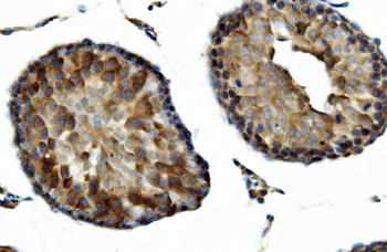
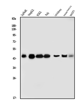
Item 1 of 8
| Tested Applications | ELISA, FC, IHC, WB |
|---|---|
| Dilution Range | Western blot, 0.25-0.5μg/ml, Human, Mouse, Rat Immunohistochemistry (Paraffin-embedded Section), 0.5-1μg/ml, Human, Mouse, Rat Flow Cytometry (Fixed), 1-3μg/1x10^6 cells, Human, Mouse, Rat ELISA, 0.1-0.5μg/ml |
| Reactivity | Human, Mouse, Rat |
Related Conjugates & Formulations
−Key Properties
−| Antibody Type | Primary Antibody |
|---|---|
| Host | Rabbit |
| Clonality | Polyclonal |
| Isotype | Rabbit IgG |
| Immunogen | E.coli-derived human DNAJA2 recombinant protein (Position: M1-N140). |
| Target | DnaJ homolog subfamily A member 2 |
| Molecular Weight | 46 kDa |
| Purification | Immunogen affinity purified. |
| Conjugation | Unconjugated |
Storage & Handling
−| Storage | Maintain refrigerated at 2-8°C for up to 2 weeks. For long term storage store at -20°C in small aliquots to prevent freeze-thaw cycles. |
|---|---|
| Form/Appearance | Lyophilized |
| Buffer/Preservatives | Each vial contains 4mg Trehalose, 0.9mg NaCl, 0.2mg Na2HPO4, 0.05mg NaN3. |
| Concentration | 500 µg/ml |
| Expiration Date | 12 months from date of receipt. |
| Disclaimer | For research use only |
Alternative Names
−CPR3; DJ3; DJA2; DNAJ; DNAJA2; DNJ3; HIRA interacting protein 4; HIRIP4; PRO3015; RDJ2
Similar Products
−- Item 1 of 2
DNAJA2 Rabbit Polyclonal Antibody [orb341230]
IF, WB
Human, Mouse, Rat
Rabbit
Polyclonal
Unconjugated
50 μl, 100 μl, 200 μl, 30 μl - Item 1 of 1
- Item 1 of 1
DNAJA2 Rabbit Polyclonal Antibody [orb2951321]
ELISA, IHC, WB
Human
Rabbit
Polyclonal
Unconjugated
50 μg, 100 μg - Item 1 of 1
DNAJA2 Rabbit Polyclonal Antibody [orb626435]
ELISA, IHC, WB
Human, Mouse
Rabbit
Polyclonal
Unconjugated
50 μg, 100 μg - Item 1 of 1
DNAJA2 Rabbit Polyclonal Antibody [orb330663]
WB
Bovine, Canine, Equine, Guinea pig, Mouse, Rabbit, Rat, Yeast, Zebrafish
Human
Rabbit
Polyclonal
Unconjugated
100 μl

Quality Guarantee
Explore bioreagents carefree to elevate your research. All our products are rigorously tested for performance. If a product does not perform as described on its datasheet, our scientific support team will provide expert troubleshooting, a prompt replacement, or a refund. For full details, please see our Terms & Conditions and Buying Guide. Contact us at support@biorbyt.com.
Quick Database Links
Gene Symbol
DnaJ homolog subfamily A member 2
UniProt
UniProt Details
− No UniProt data available
Protocol Information
WB
Western Blot (IB, immunoblot)
IHC
Immunohistochemistry
FC
Flow Cytometry
ELISA
Enzyme-linked Immunosorbent Assay (EIA)
Available Sizes
Select a size below
Free Secondary Antibody (20 ul)0/0
Please add an antibody product to your cart first.