You have no items in your shopping cart.
Description
Research Area
Cell Biology
Images & Validation
−Item 1 of 6
| Tested Applications | FC, ICC, WB |
|---|---|
| Reactivity | Bovine, Gallus, Human, Mouse, Plant, Porcine, Protozoa, Rat |
| Application Notes |
Key Properties
−| Antibody Type | Primary Antibody |
|---|---|
| Clonality | Monoclonal |
| Isotype | Mouse IgG1 |
| Clone No. | TU-30 |
| Immunogen | C-terminal peptide of gamma-tubulin counjugated to KLH. |
| Target | gamma-Tubulin |
| Purification | Purified by protein-A affinity chromatography. |
| Conjugation | Unconjugated |
Storage & Handling
−| Storage | Maintain refrigerated at 2-8°C for up to 2 weeks. For long term storage store at -20°C in small aliquots to prevent freeze-thaw cycles. |
|---|---|
| Buffer/Preservatives | Phosphate buffered saline (PBS), pH 7.4, 15 mM sodium azide |
| Concentration | 1 mg/ml |
| Expiration Date | 12 months from date of receipt. |
| Disclaimer | For research use only |
Alternative Names
−TUBG
Similar Products
−- Item 1 of 4
GCP5 rabbit pAb Antibody [orb765287]
ELISA, IF, IHC, WB
Human, Mouse, Rat
Polyclonal
Unconjugated
100 μl - Item 1 of 4
GCP6 rabbit pAb Antibody [orb765288]
ELISA, IF, IHC, WB
Human, Mouse, Rat
Polyclonal
Unconjugated
100 μl, 50 μl - Item 1 of 4
Gamma Tubulin Mouse Monoclonal Antibody [orb499656]
FC, IF, IHC-Fr, IHC-P, WB
Mouse, Rat
Human, Mouse, Rat
Mouse
Monoclonal
Unconjugated
200 μg, 50 μl, 100 μl, 200 μl - Item 1 of 3
Gamma Tubulin (6C12) Mouse mAb Antibody [orb763568]
IHC, WB
Human, Mouse, Rat
Monoclonal
Unconjugated
100 μl, 50 μl - Item 1 of 3
Quality Guarantee
Explore bioreagents carefree to elevate your research. All our products are rigorously tested for performance. If a product does not perform as described on its datasheet, our scientific support team will provide expert troubleshooting, a prompt replacement, or a refund. For full details, please see our Terms & Conditions and Buying Guide. Contact us at support@biorbyt.com.
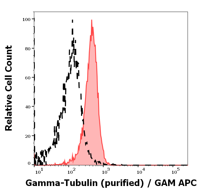
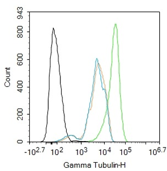
Separation of MCF-7 cells stained using anti-gamma-Tubulin (TU-30) purified antibody (concentration in sample 9 µg/ml, GAM APC, red-filled) from MCF-7 cells unstained by primary antibody (GAM APC, black-dashed) in flow cytometry analysis (intracellular staining).
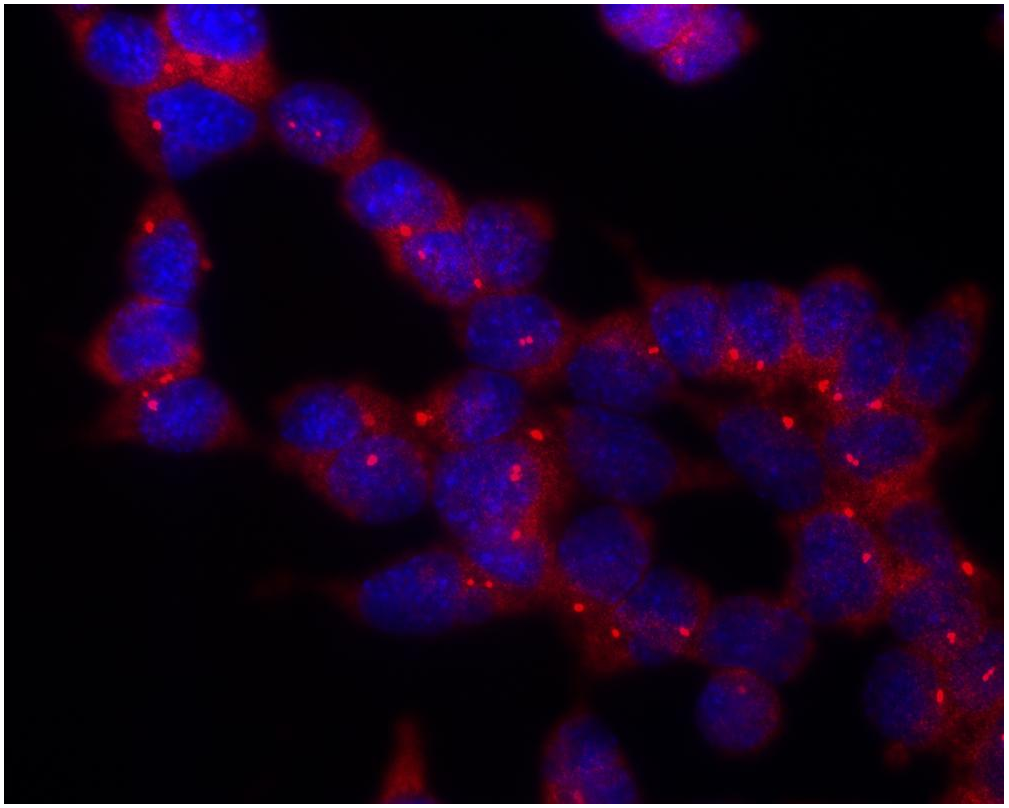
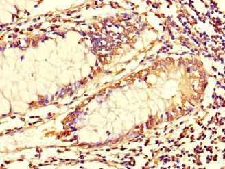
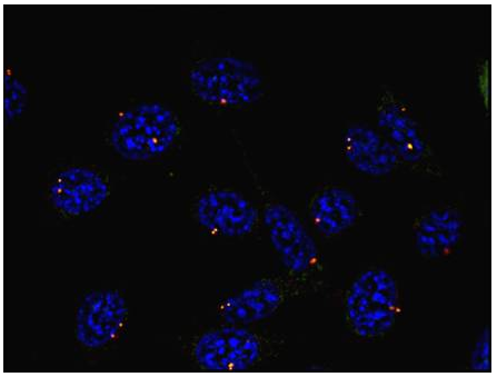
Immunocytochemistry staining of P19X1 mouse embryonal carcinoma cell line using anti-gamma-tubulin (TU-30) (detection by secondary antibody Goat anti-mouse Cy3). Nuclei were stained with DAPI (blue).
Immunocytochemistry staining of murine fibroblasts using anti-gamma-tubulin (TU-30; direct conjugate with Dyomics 547, red). Nuclei were stained with DAPI (blue).
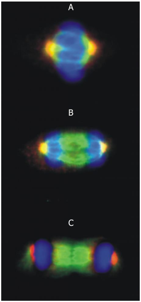
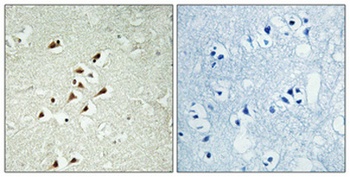
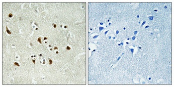
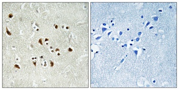
Immunocytochemistry staining of microtubular networks in 3T3 mouse fibroblasts. A - metaphase; B - anaphase; C - telophase. Gamma-tubulin (red) stained with anti-gamma-tubulin (TU-30), alpha-tubulin (green) with polyclonal anti-alpha-tubulin antibody and nuclei with DAPI (blue).
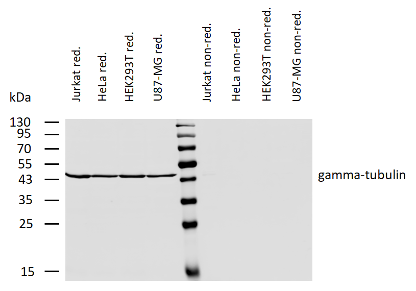
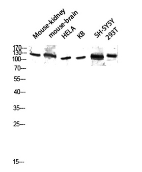
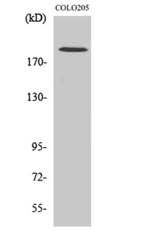
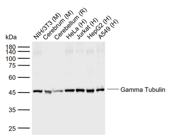
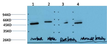
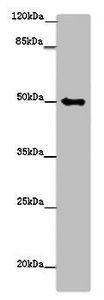
Western blotting analysis of human gamma-tubulin using mouse monoclonal antibody TU-30 on lysates of various cell lines under reducing and non-reducing conditions. Nitrocellulose membrane was probed with 2 µg/ml of mouse anti-gamma-tubulin monoclonal antibody followed by IRDye800-conjugated anti-mouse secondary antibody. A specific band was detected for gamma-tubulin at approximately 46 kDa.
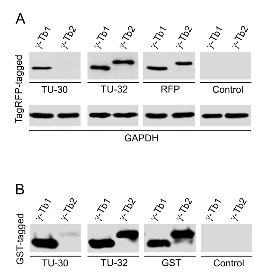
Western blotting analysis of differential reactivity of monoclonal antibodies to γ-tubulin with human γ-tubulin isotypes. (A) Immunoblots of total cell lysates from SH-SY5Y cells, expressing TagRFP-tagged human γ-tubulin 1 (γ-Tb1) or γ-tubulin 2 (γ-Tb2), probed with Abs to γ-tubulin (TU-30, TU-32), TagRFP (RFP) and GAPDH. In control samples, only secondary anti-mouse Ab was applied. (B) Immunoblots of immobilized GST-tagged human C-terminal regions (a.a. 362-451) of γ-Tb1 or γ-Tb2 probed with Abs to γ-tubulin (TU-30, TU-32) and GST. In control samples, only secondary anti-mouse Ab was applied.
Documents Download
Datasheet
Product Information
Request a Document
Protocol Information
WB
Western Blot (IB, immunoblot)
FC
Flow Cytometry
ICC
Immunocytochemistry
gamma-Tubulin Antibody (orb44548)

- 0.0
Based on 0 reviews
Participating in our Biorbyt product reviews program enables you to support fellow scientists by sharing your firsthand experience with our products.
Login to Submit a ReviewAvailable Sizes
Select a size below
Free Secondary Antibody (20 ul)0/0
Please add an antibody product to your cart first.