You have no items in your shopping cart.
Description
Research Area
Cell Biology
Images & Validation
−Item 1 of 3
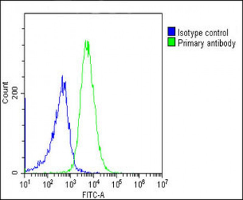
| Tested Applications | IHC-Fr, IHC-P, WB |
|---|---|
| Reactivity | Human |
| Application Notes |
Key Properties
−| Antibody Type | Primary Antibody |
|---|---|
| Clonality | Monoclonal |
| Isotype | Mouse IgG1 |
| Clone No. | MEM-G/1 |
| Immunogen | Denatured bacterially expressed recombinant human HLA-G heavy chain. |
| Target | HLA-G |
| Purification | Purified by protein-A affinity chromatography. |
| Conjugation | Unconjugated |
Storage & Handling
−| Storage | Maintain refrigerated at 2-8°C for up to 2 weeks. For long term storage store at -20°C in small aliquots to prevent freeze-thaw cycles. |
|---|---|
| Buffer/Preservatives | Phosphate buffered saline (PBS), pH 7.4, 15 mM sodium azide |
| Concentration | 1 mg/ml |
| Expiration Date | 12 months from date of receipt. |
| Disclaimer | For research use only |
Similar Products
−- Item 1 of 5
- Item 1 of 6
- Item 1 of 1
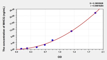
Human Major Histocompatibility Complex Class I G (MHCG) ELISA Kit [orb776093]
Human
0.38-24 ng/mL
0.17 ng/mL
48 T, 96 T - Item 1 of 3
- Item 1 of 3
HLA-G Antibody [orb2309990]
IHC, WB
Human
Mouse
Monoclonal
Unconjugated
20 μg, 100 μg (without BSA and Azide), 100 μg


Quality Guarantee
Explore bioreagents carefree to elevate your research. All our products are rigorously tested for performance. If a product does not perform as described on its datasheet, our scientific support team will provide expert troubleshooting, a prompt replacement, or a refund. For full details, please see our Terms & Conditions and Buying Guide. Contact us at support@biorbyt.com.
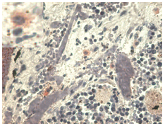
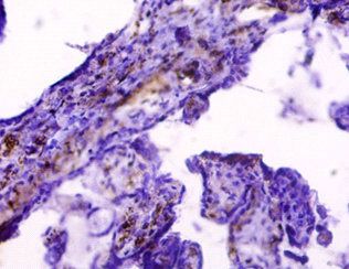
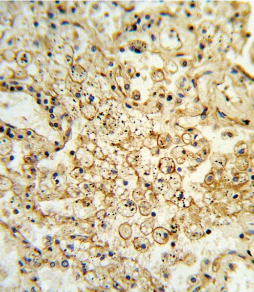
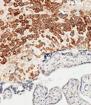
Immunohistochemistry staining with anti-human HLA-G (MEM-G/1) - pulmonary disseases (paraffin-embedded sections). The antibody MEM-G/1 stains infiltrating macrophages in pulmonary diseases. In the top left corner see the detail of macrophage.
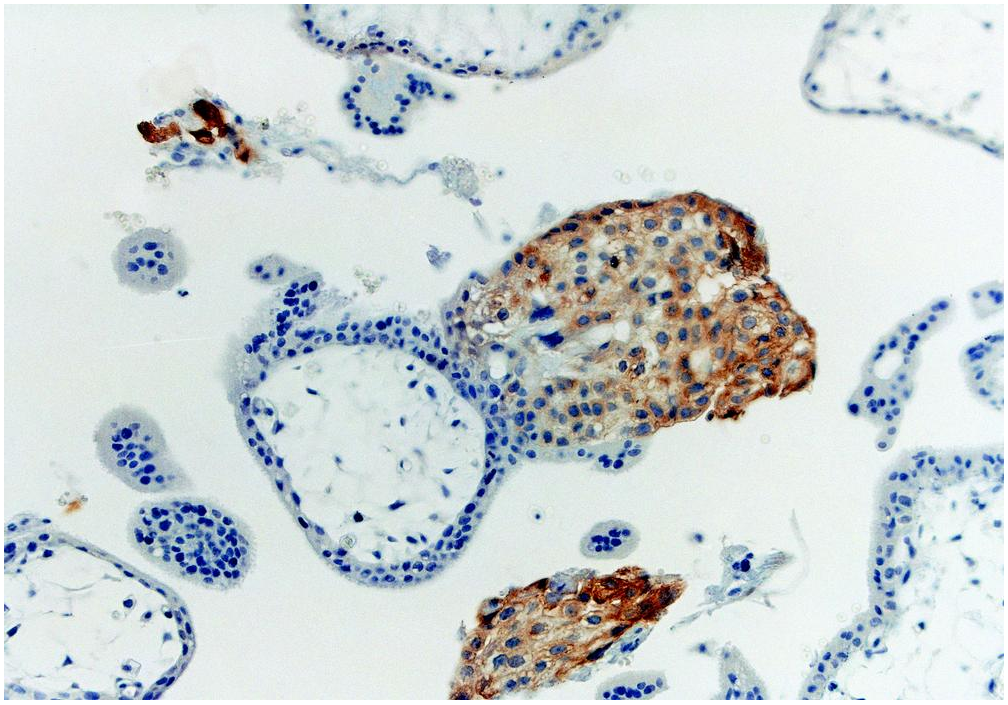
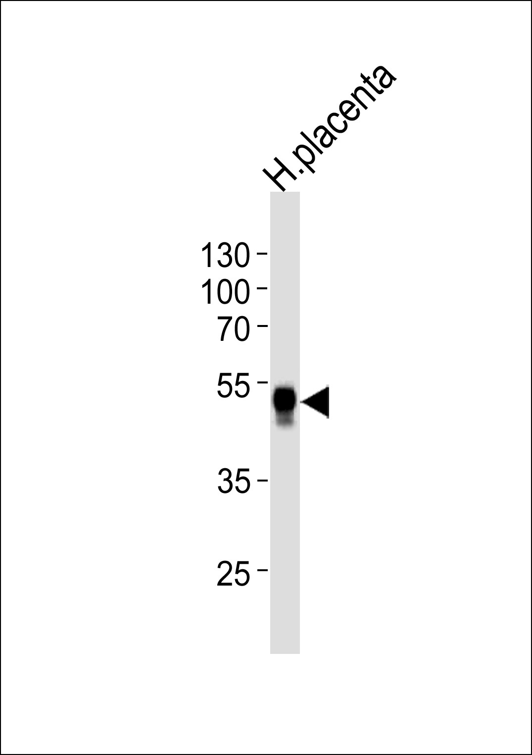
Immunohistochemistry staining with anti-human HLA-G (MEM-G/1) of first trimester placenta (paraffin-embedded sections).
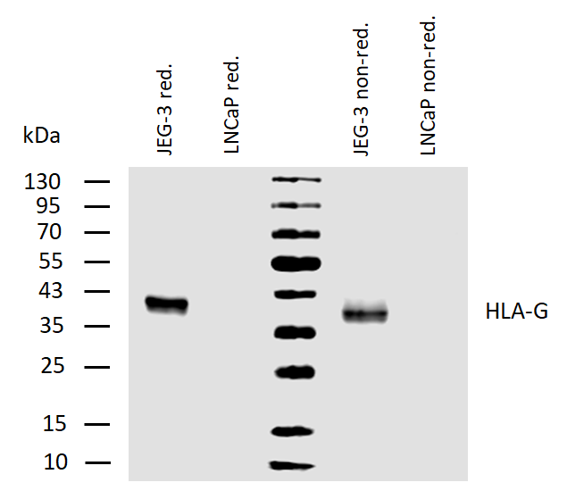
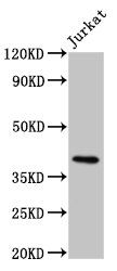
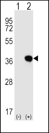
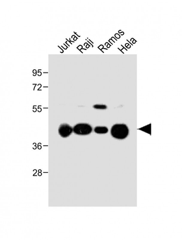
Western blotting analysis of human HLA-G using mouse monoclonal antibody MEM-G/1 on lysates of JEG-3 cell line and LNCaP cell line (negative control) under reducing and non-reducing conditions. Nitrocellulose membrane was probed with 2 µg/ml of mouse monoclonal antibody anti-HLA-G followed by IRDye800-conjugated anti-mouse secondary antibody. HLA-G was detected at approximately 40 kDa.
Documents Download
Datasheet
Product Information
Request a Document
Protocol Information
WB
Western Blot (IB, immunoblot)
IHC-P
Immunohistochemistry Paraffin
IHC-Fr
Immunohistochemistry Frozen
HLA-G Antibody (orb44426)

- 0.0
Based on 0 reviews
Participating in our Biorbyt product reviews program enables you to support fellow scientists by sharing your firsthand experience with our products.
Login to Submit a ReviewAvailable Sizes
Select a size below
Free Secondary Antibody (20 ul)0/0
Please add an antibody product to your cart first.