You have no items in your shopping cart.
Description
Research Area
Immunology & Inflammation
Images & Validation
−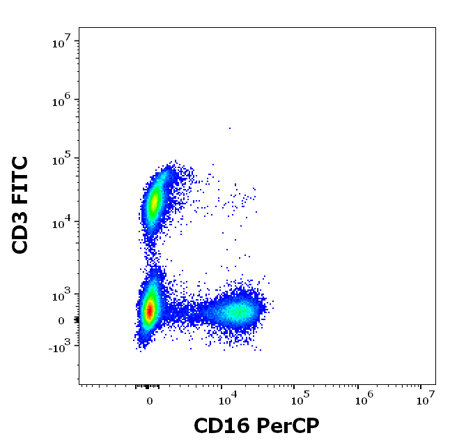
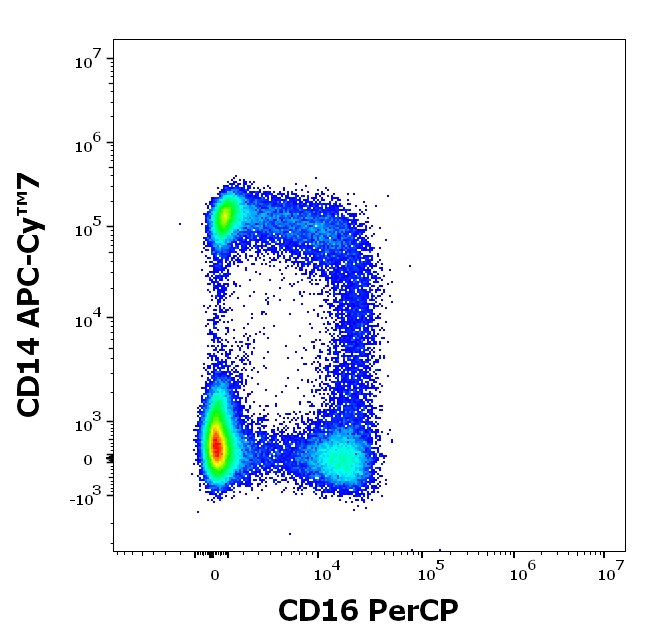


Item 1 of 4
| Tested Applications | FC |
|---|---|
| Reactivity | Human, Primate |
| Application Notes |
Key Properties
−| Antibody Type | Primary Antibody |
|---|---|
| Clonality | Monoclonal |
| Isotype | Mouse IgG1 kappa |
| Clone No. | 3G8 |
| Immunogen | Human neutrophils |
| Target | CD16 |
| Purification | Purified antibody is conjugated with activated Peridinin-Chlorophyll Protein (PerCP) under optimum conditions and unconjugated antibody and free fluorochrome are removed by size-exclusion chromatography. |
| Conjugation | PerCP |
Storage & Handling
−| Storage | Store at 2-8°C. Protect from prolonged exposure to light. Do not freeze. |
|---|---|
| Buffer/Preservatives | Stabilizing phosphate buffered saline (PBS), pH 7.4, 15 mM sodium azide |
| Expiration Date | 12 months from date of receipt. |
| Disclaimer | For research use only |
Alternative Names
−FcgammaRIII, IGFR3, FCRIII
Similar Products
−
CD16 Mouse Monoclonal Antibody (PerCP) [orb2972115]
ELISA, FC, IF
Human
Mouse
Monoclonal
PerCP
100 T, 50 THuman CD16 Mouse Monoclonal Antibody (PerCP) [orb1184659]
FC
Human
Mouse
Monoclonal
PerCP
25 T, 50 T, 100 TCD16 Mouse Monoclonal Antibody (PerCP) [orb1293516]
FC, IF
Human
Mouse
Monoclonal
PerCP
100 assay, 25 assaymouse CD16/32 Rat mAb, PerCP-Cy5.5 conjugated [orb2369991]
FC
Mouse
Mouse
Rat
Monoclonal
PerCP/Cy5.5
100 μl
Quality Guarantee
Explore bioreagents carefree to elevate your research. All our products are rigorously tested for performance. If a product does not perform as described on its datasheet, our scientific support team will provide expert troubleshooting, a prompt replacement, or a refund. For full details, please see our Terms & Conditions and Buying Guide. Contact us at support@biorbyt.com.
Available Sizes
Select a size below
Free Secondary Antibody (20 ul)0/0
Please add an antibody product to your cart first.