You have no items in your shopping cart.
Description
Research Area
Epigenetics, Immunology
Images & Validation
−Item 1 of 3
| Tested Applications | FC |
|---|---|
| Reactivity | Human |
| Application Notes |
Key Properties
−| Antibody Type | Primary Antibody |
|---|---|
| Clonality | Monoclonal |
| Isotype | Mouse IgG1 |
| Clone No. | HI149 |
| Immunogen | Human thymocytes |
| Target | CD1a |
| Purification | Purified antibody is conjugated with R-phycoerythrin (PE) under optimum conditions. Unconjugated antibody and free fluorochrome are removed by size-exclusion chromatography. |
| Conjugation | PE |
Storage & Handling
−| Storage | Store at 2-8°C. Protect from prolonged exposure to light. Do not freeze. |
|---|---|
| Buffer/Preservatives | Stabilizing phosphate buffered saline (PBS), pH 7.4, 15 mM sodium azide |
| Expiration Date | 12 months from date of receipt. |
| Disclaimer | For research use only |
Alternative Names
−T6, Leu-6, HTA1, FCB6
Similar Products
−- Item 1 of 1
CD1a Mouse Monoclonal Antibody (PE/Cy7) [orb1293549]
FC, IF
Human
Mouse
Monoclonal
PE/Cy7
25 assay, 100 assayHuman CD1a Mouse Monoclonal Antibody (PE) [orb1184662]
FC
Human
Human
Mouse
Monoclonal
PE
25 T, 100 T, 50 T
Quality Guarantee
Explore bioreagents carefree to elevate your research. All our products are rigorously tested for performance. If a product does not perform as described on its datasheet, our scientific support team will provide expert troubleshooting, a prompt replacement, or a refund. For full details, please see our Terms & Conditions and Buying Guide. Contact us at support@biorbyt.com.
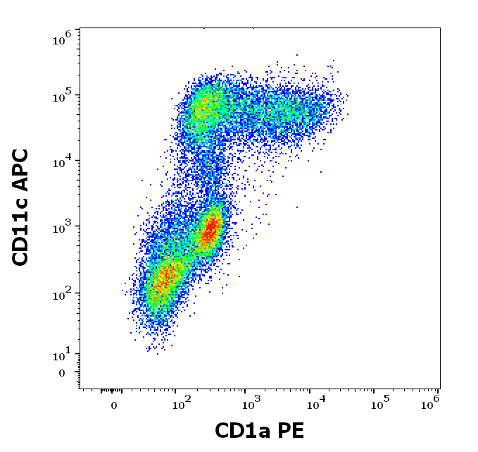
Flow cytometry multicolor surface staining of human stimulated (GM-CSF + IL-4) peripheral blood monocytes stained using anti-human CD1a (HI149) PE antibody (20 µl reagent per milion cells in 100 µl of cell suspension) and anti-human CD11c (BU15) APC antibody (10 µl reagent per milion cells in 100 µl of cell suspension).
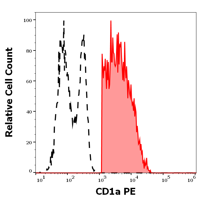
Separation of human CD1a positive CD11c positive dendritic cells differentiated upon monocyte stimulation (GM-CSF + IL-4) (red-filled) from CD11c negative CD1a negative events (black-dashed) in flow cytometry analysis (surface staining) of human stimulated (GM-CSF + IL-4) peripheral blood monocytes stained using CD1a (HI149) PE antibody (20 µl reagent per milion cells in 100 µl of cell suspension).

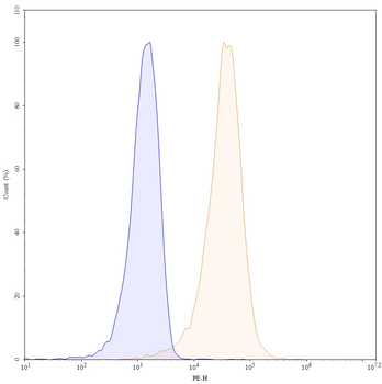
Flow cytometry surface staining pattern of human stimulated (GM-CSF + IL-4) peripheral blood monocytes stained using anti-human CD1a (HI149) PE antibody (20 µl reagent per milion cells in 100 µl of cell suspension).
Documents Download
Datasheet
Product Information
Request a Document
CD1a Antibody (PE) (orb43816)

- 0.0
Based on 0 reviews
Participating in our Biorbyt product reviews program enables you to support fellow scientists by sharing your firsthand experience with our products.
Login to Submit a ReviewAvailable Sizes
Select a size below
Free Secondary Antibody (20 ul)0/0
Please add an antibody product to your cart first.