You have no items in your shopping cart.
Description
Research Area
Cancer Biology, Cardiovascular Research, Cell Biology, Immunology & Inflammation, Metabolism Research
Images & Validation
−
Item 1 of 3
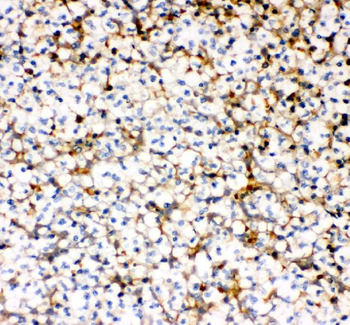
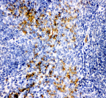
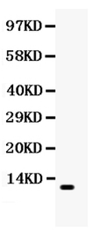
| Tested Applications | ELISA, IHC, WB |
|---|---|
| Dilution Range | Western blot, 0.1-0.5μg/ml, Human Immunohistochemistry (Paraffin-embedded Section), 0.5-1μg/ml, Human ELISA, 0.1-0.5μg/ml |
| Reactivity | Human |
Related Conjugates & Formulations
−Key Properties
−| Antibody Type | Primary Antibody |
|---|---|
| Host | Rabbit |
| Clonality | Polyclonal |
| Isotype | Rabbit IgG |
| Immunogen | E. coli-derived human MCP-1 recombinant protein (Position: Q24-T99). Human MCP-1 shares 60.9% and 59.4% amino acid (aa) sequence identity with mouse and rat MCP-1, respectively. |
| Target | C-C motif chemokine 2 |
| Molecular Weight | 11 kDa |
| Purification | Immunogen affinity purified. |
| Conjugation | Unconjugated |
Storage & Handling
−| Storage | Maintain refrigerated at 2-8°C for up to 2 weeks. For long term storage store at -20°C in small aliquots to prevent freeze-thaw cycles. |
|---|---|
| Form/Appearance | Lyophilized |
| Buffer/Preservatives | Each vial contains antibody formulated with stabilizing components, 0.9 mg NaCl, 0.2 mg Na2HPO4, and 0.05 mg NaN3. *This antibody is supplied in a stabilized formulation. Compatibility with conjugation reactions depends on the chemistry of the conjugation method used. For conjugation methods that are not compatible with the stabilizing components present in this formulation, a carrier-free antibody format is required. |
| Concentration | Adding 0.2 ml of distilled water will yield a concentration of 500 μg/ml. |
| Expiration Date | 12 months from date of receipt. |
| Disclaimer | For research use only |
Alternative Names
−C-C motif chemokine 2; HC11; Monocyte chemoattractant protein 1; Monocyte chemotactic and activating factor; MCAF; Monocyte chemotactic protein 1; MCP-1; Monocyte secretory protein JE; Small-inducible cytokine A2; CCL2; MCP1, SCYA2
Similar Products
−
MCP1/CCL2 Rabbit Polyclonal Antibody (Carrier-free) [orb3076785]
ELISA, IHC, WB
Human
Rabbit
Polyclonal
Carrier-free
100 μg
Quality Guarantee
Explore bioreagents carefree to elevate your research. All our products are rigorously tested for performance. If a product does not perform as described on its datasheet, our scientific support team will provide expert troubleshooting, a prompt replacement, or a refund. For full details, please see our Terms & Conditions and Buying Guide. Contact us at support@biorbyt.com.
Quick Database Links
Gene Symbol
C-C motif chemokine 2
UniProt
UniProt Details
− No UniProt data available
Protocol Information
WB
Western Blot (IB, immunoblot)
IHC
Immunohistochemistry
ELISA
Enzyme-linked Immunosorbent Assay (EIA)
Available Sizes
Select a size below
Free Secondary Antibody (20 ul)0/0
Please add an antibody product to your cart first.