You have no items in your shopping cart.
Myc-tag Antibody
SKU: orb323058
Description
Images & Validation
−Item 1 of 3
| Tested Applications | IF, IP, WB |
|---|---|
| Dilution Range | WB: 1:2000-1:5000, IF/ICC: 1:200-1:500, IP: 1:100-1:200 |
Key Properties
−| Antibody Type | Primary Antibody |
|---|---|
| Host | Mouse |
| Clonality | Monoclonal |
| Immunogen | KLH-conjugated synthetic peptide encompassing a sequence of Myc-tag. The exact sequence is proprietary. |
| Purification | Affinity chromatography |
| Conjugation | Unconjugated |
Storage & Handling
−| Storage | Maintain refrigerated at 2-8°C for up to 2 weeks. For long term storage store at -20°C in small aliquots to prevent freeze-thaw cycles. |
|---|---|
| Form/Appearance | Liquid in 0.42% Potassium phosphate, 0.87% Sodium chloride, pH 7.3, 30% glycerol, and 0.01% sodium azide. |
| Expiration Date | 12 months from date of receipt. |
| Disclaimer | For research use only |
Similar Products
−- Item 1 of 3
- Item 1 of 4
Rabbit Myc Tag Recombinant Monoclonal Antibody [orb1530834]
ELISA, ICC, IP, WB
Rabbit
Recombinant
Unconjugated
100 μl - Item 1 of 2
- Item 1 of 2
Anti-c-myc epitope tag [9E10] [orb256348]
IF, IHC-Fr, IHC-P, IP, WB
Human
Mouse
Monoclonal
Unconjugated
0.2 mg - Item 1 of 2
Anti-c-myc epitope tag [9E10] [orb256349]
IF, IHC-Fr, IHC-P, IP, WB
Human
Mouse
Monoclonal
Unconjugated
0.1 mg
![Anti-c-myc epitope tag [9E10]](/images/pub/media/catalog/product/NewWebsite/35/orb256348_1.png)
![Anti-c-myc epitope tag [9E10]](/images/pub/media/catalog/product/NewWebsite/35/orb256348_2.png)
![Anti-c-myc epitope tag [9E10]](/images/pub/media/catalog/product/NewWebsite/35/orb256349_1.png)
![Anti-c-myc epitope tag [9E10]](/images/pub/media/catalog/product/NewWebsite/35/orb256349_2.png)
Quality Guarantee
Explore bioreagents carefree to elevate your research. All our products are rigorously tested for performance. If a product does not perform as described on its datasheet, our scientific support team will provide expert troubleshooting, a prompt replacement, or a refund. For full details, please see our Terms & Conditions and Buying Guide. Contact us at support@biorbyt.com.
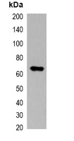
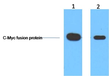
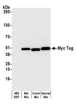
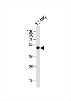
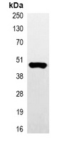
Western blot analysis of over-expressed Myc-tagged protein in 293T cell lysate. (Predicted band size: \; Observed band size: \)
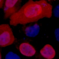
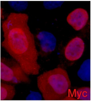
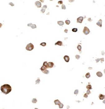
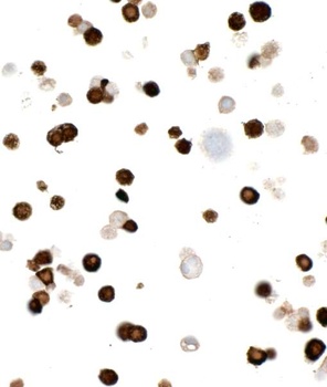
Immunofluorescent analysis of Myc-tag staining in 293T cells transfected with a Myc-tag protein. Formalin-fixed cells were permeabilized with 0.1% Triton X-100 in TBS for 5-10 minutes and blocked with 3% BSA-PBS for 30 minutes at room temperature. Cells were probed with the primary antibody in 3% BSA-PBS and incubated overnight at 4 °C in a humidified chamber. Cells were washed with PBST and incubated with a DyLight 594-conjugated secondary antibody (red) in PBS at room temperature in the dark. DAPI was used to stain the cell nuclei (blue).
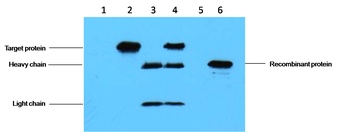
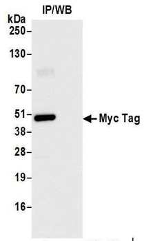
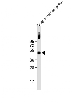
Immunoprecipitation of Myc-tagged protein from HEK293T cells transfected with vector overexpressing Myc tag, using Anti-Myc-tag Antibody.
Documents Download
Datasheet
Product Information
Request a Document
Protocol Information
WB
Western Blot (IB, immunoblot)
IF
Immunofluorescence
IP
Immunoprecipitation
Myc-tag Antibody (orb323058)

- 0.0
Based on 0 reviews
Participating in our Biorbyt product reviews program enables you to support fellow scientists by sharing your firsthand experience with our products.
Login to Submit a ReviewAvailable Sizes
Select a size below