You have no items in your shopping cart.
Description
Research Area
Images & Validation
−| Tested Applications | ELISA, FC, IHC, WB |
|---|---|
| Dilution Range | Western blot, 0.25-0.5μg/ml, Human, Monkey, Mouse, Rat Immunohistochemistry (Paraffin-embedded Section), 0.5-1μg/ml, Human, Mouse, Rat Flow Cytometry (Fixed), 1-3μg/1x10^6 cells, Human ELISA, 0.1-0.5μg/ml |
| Reactivity | Human, Monkey, Mouse, Rat |
Related Conjugates & Formulations
−Key Properties
−| Antibody Type | Primary Antibody |
|---|---|
| Host | Rabbit |
| Clonality | Polyclonal |
| Isotype | Rabbit IgG |
| Immunogen | E.coli-derived human Osteoprotegerin/TNFRSF11B recombinant protein (Position: Q247-R296). |
| Target | Tumor necrosis factor receptor superfamily member 11B |
| Molecular Weight | 55 kDa |
| Purification | Immunogen affinity purified. |
| Conjugation | Unconjugated |
Storage & Handling
−| Storage | Maintain refrigerated at 2-8°C for up to 2 weeks. For long term storage store at -20°C in small aliquots to prevent freeze-thaw cycles. |
|---|---|
| Form/Appearance | Lyophilized |
| Buffer/Preservatives | Each vial contains 4mg Trehalose, 0.9mg NaCl, 0.2mg Na2HPO4, 0.05mg NaN3. |
| Concentration | 500 µg/ml |
| Expiration Date | 12 months from date of receipt. |
| Disclaimer | For research use only |
Alternative Names
−Similar Products
−TNFRSF11B Antibody (Center) [orb1437242]
IF, IHC-P, WB
Human
Rabbit
Polyclonal
Unconjugated
50 μl, 100 μlTNFRSF11B Rabbit Polyclonal Antibody [orb2955937]
ELISA, IHC, WB
Bovine, Human, Mouse, Rat
Rabbit
Polyclonal
Unconjugated
50 μg, 100 μgOsteoprotegerin/TNFRSF11B Rabbit Polyclonal Antibody (Fluoro550) [orb3116504]
Human, Monkey, Mouse, Rat
Rabbit
Polyclonal
Fluoro550
100 μgOsteoprotegerin/TNFRSF11B Rabbit Polyclonal Antibody (Fluoro647) [orb3116502]
Human, Monkey, Mouse, Rat
Rabbit
Polyclonal
Fluoro647
100 μgOsteoprotegerin/TNFRSF11B Rabbit Polyclonal Antibody (Fluoro594) [orb3116503]
Human, Monkey, Mouse, Rat
Rabbit
Polyclonal
Fluoro594
100 μg

Quality Guarantee
Explore bioreagents carefree to elevate your research. All our products are rigorously tested for performance. If a product does not perform as described on its datasheet, our scientific support team will provide expert troubleshooting, a prompt replacement, or a refund. For full details, please see our Terms & Conditions and Buying Guide. Contact us at support@biorbyt.com.

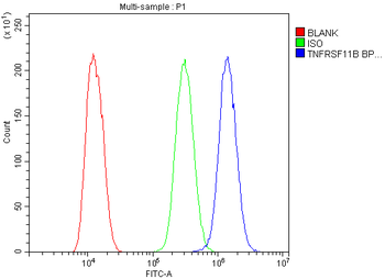
Flow Cytometry analysis of U20S cells using anti-TNFRSF11B antibody. Overlay histogram showing U20S cells (Blue line). To facilitate intracellular staining, cells were fixed with 4% paraformaldehyde and permeabilized with permeabilization buffer. The cells were blocked with 10% normal goat serum. And then incubated with rabbit anti-TNFRSF11B Antibody (1 µg/1x10^6 cells) for 30 min at 20°C. DyLight®488 conjugated goat anti-rabbit IgG (5-10 µg/1x10^6 cells) was used as secondary antibody for 30 minutes at 20°C. Isotype control antibody (Green line) was rabbit IgG (1 µg/1x10^6) used under the same conditions. Unlabelled sample without incubation with primary antibody and secondary antibody (Red line) was used as a blank control.

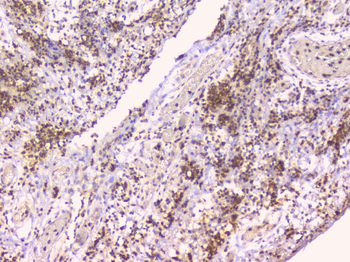
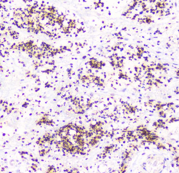
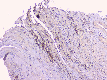
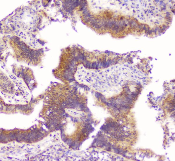
IHC analysis of TNFRSF11B using anti-TNFRSF11B antibody. TNFRSF11B was detected in paraffin-embedded section of human appendicitis tissues. Heat mediated antigen retrieval was performed in citrate buffer (pH6, epitope retrieval solution) for 20 mins. The tissue section was blocked with 10% goat serum. The tissue section was then incubated with 1 µg/ml rabbit anti-TNFRSF11B Antibody overnight at 4°C. Biotinylated goat anti-rabbit IgG was used as secondary antibody and incubated for 30 minutes at 37°C. The tissue section was developed using Strepavidin-Biotin-Complex (SABC) with DAB as the chromogen.

IHC analysis of TNFRSF11B using anti-TNFRSF11B antibody. TNFRSF11B was detected in paraffin-embedded section of human appendicitis tissues. Heat mediated antigen retrieval was performed in citrate buffer (pH6, epitope retrieval solution) for 20 mins. The tissue section was blocked with 10% goat serum. The tissue section was then incubated with 1 µg/ml rabbit anti-TNFRSF11B Antibody overnight at 4°C. Biotinylated goat anti-rabbit IgG was used as secondary antibody and incubated for 30 minutes at 37°C. The tissue section was developed using Strepavidin-Biotin-Complex (SABC) with DAB as the chromogen.

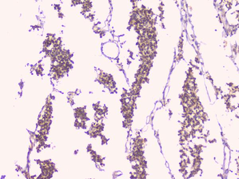
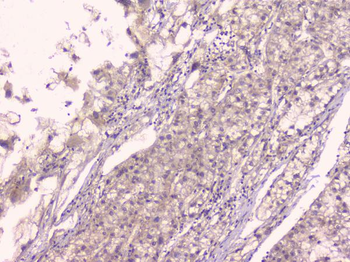
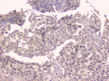
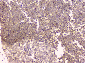
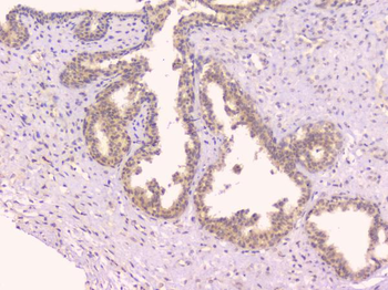
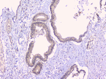
IHC analysis of TNFRSF11B using anti-TNFRSF11B antibody. TNFRSF11B was detected in paraffin-embedded section of human Cholangiocarcinoma tissues. Heat mediated antigen retrieval was performed in citrate buffer (pH6, epitope retrieval solution) for 20 mins. The tissue section was blocked with 10% goat serum. The tissue section was then incubated with 1 µg/ml rabbit anti-TNFRSF11B Antibody overnight at 4°C. Biotinylated goat anti-rabbit IgG was used as secondary antibody and incubated for 30 minutes at 37°C. The tissue section was developed using Strepavidin-Biotin-Complex (SABC) with DAB as the chromogen.

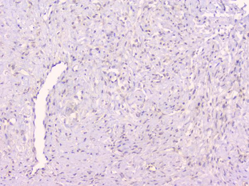
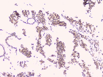
IHC analysis of TNFRSF11B using anti-TNFRSF11B antibody. TNFRSF11B was detected in paraffin-embedded section of human endometrial carcinoma tissues. Heat mediated antigen retrieval was performed in citrate buffer (pH6, epitope retrieval solution) for 20 mins. The tissue section was blocked with 10% goat serum. The tissue section was then incubated with 1 µg/ml rabbit anti-TNFRSF11B Antibody overnight at 4°C. Biotinylated goat anti-rabbit IgG was used as secondary antibody and incubated for 30 minutes at 37°C. The tissue section was developed using Strepavidin-Biotin-Complex (SABC) with DAB as the chromogen.

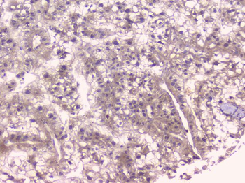
IHC analysis of TNFRSF11B using anti-TNFRSF11B antibody. TNFRSF11B was detected in paraffin-embedded section of human glioma tissues. Heat mediated antigen retrieval was performed in citrate buffer (pH6, epitope retrieval solution) for 20 mins. The tissue section was blocked with 10% goat serum. The tissue section was then incubated with 1 µg/ml rabbit anti-TNFRSF11B Antibody overnight at 4°C. Biotinylated goat anti-rabbit IgG was used as secondary antibody and incubated for 30 minutes at 37°C. The tissue section was developed using Strepavidin-Biotin-Complex (SABC) with DAB as the chromogen.

IHC analysis of TNFRSF11B using anti-TNFRSF11B antibody. TNFRSF11B was detected in paraffin-embedded section of human glioma tissues. Heat mediated antigen retrieval was performed in citrate buffer (pH6, epitope retrieval solution) for 20 mins. The tissue section was blocked with 10% goat serum. The tissue section was then incubated with 1 µg/ml rabbit anti-TNFRSF11B Antibody overnight at 4°C. Biotinylated goat anti-rabbit IgG was used as secondary antibody and incubated for 30 minutes at 37°C. The tissue section was developed using Strepavidin-Biotin-Complex (SABC) with DAB as the chromogen.

IHC analysis of TNFRSF11B using anti-TNFRSF11B antibody. TNFRSF11B was detected in paraffin-embedded section of human liver cancer tissues. Heat mediated antigen retrieval was performed in citrate buffer (pH6, epitope retrieval solution) for 20 mins. The tissue section was blocked with 10% goat serum. The tissue section was then incubated with 1 µg/ml rabbit anti-TNFRSF11B Antibody overnight at 4°C. Biotinylated goat anti-rabbit IgG was used as secondary antibody and incubated for 30 minutes at 37°C. The tissue section was developed using Strepavidin-Biotin-Complex (SABC) with DAB as the chromogen.

IHC analysis of TNFRSF11B using anti-TNFRSF11B antibody. TNFRSF11B was detected in paraffin-embedded section of human liver cancer tissues. Heat mediated antigen retrieval was performed in citrate buffer (pH6, epitope retrieval solution) for 20 mins. The tissue section was blocked with 10% goat serum. The tissue section was then incubated with 1 µg/ml rabbit anti-TNFRSF11B Antibody overnight at 4°C. Biotinylated goat anti-rabbit IgG was used as secondary antibody and incubated for 30 minutes at 37°C. The tissue section was developed using Strepavidin-Biotin-Complex (SABC) with DAB as the chromogen.

IHC analysis of TNFRSF11B using anti-TNFRSF11B antibody. TNFRSF11B was detected in paraffin-embedded section of human Lung cancer tissues. Heat mediated antigen retrieval was performed in citrate buffer (pH6, epitope retrieval solution) for 20 mins. The tissue section was blocked with 10% goat serum. The tissue section was then incubated with 1 µg/ml rabbit anti-TNFRSF11B Antibody overnight at 4°C. Biotinylated goat anti-rabbit IgG was used as secondary antibody and incubated for 30 minutes at 37°C. The tissue section was developed using Strepavidin-Biotin-Complex (SABC) with DAB as the chromogen.

IHC analysis of TNFRSF11B using anti-TNFRSF11B antibody. TNFRSF11B was detected in paraffin-embedded section of human Lung cancer tissues. Heat mediated antigen retrieval was performed in citrate buffer (pH6, epitope retrieval solution) for 20 mins. The tissue section was blocked with 10% goat serum. The tissue section was then incubated with 1 µg/ml rabbit anti-TNFRSF11B Antibody overnight at 4°C. Biotinylated goat anti-rabbit IgG was used as secondary antibody and incubated for 30 minutes at 37°C. The tissue section was developed using Strepavidin-Biotin-Complex (SABC) with DAB as the chromogen.

IHC analysis of TNFRSF11B using anti-TNFRSF11B antibody. TNFRSF11B was detected in paraffin-embedded section of human Lung cancer tissues. Heat mediated antigen retrieval was performed in citrate buffer (pH6, epitope retrieval solution) for 20 mins. The tissue section was blocked with 10% goat serum. The tissue section was then incubated with 1 µg/ml rabbit anti-TNFRSF11B Antibody overnight at 4°C. Biotinylated goat anti-rabbit IgG was used as secondary antibody and incubated for 30 minutes at 37°C. The tissue section was developed using Strepavidin-Biotin-Complex (SABC) with DAB as the chromogen.

IHC analysis of TNFRSF11B using anti-TNFRSF11B antibody. TNFRSF11B was detected in paraffin-embedded section of human mammary cancer tissues. Heat mediated antigen retrieval was performed in citrate buffer (pH6, epitope retrieval solution) for 20 mins. The tissue section was blocked with 10% goat serum. The tissue section was then incubated with 1 µg/ml rabbit anti-TNFRSF11B Antibody overnight at 4°C. Biotinylated goat anti-rabbit IgG was used as secondary antibody and incubated for 30 minutes at 37°C. The tissue section was developed using Strepavidin-Biotin-Complex (SABC) with DAB as the chromogen.

IHC analysis of TNFRSF11B using anti-TNFRSF11B antibody. TNFRSF11B was detected in paraffin-embedded section of human mammary cancer tissues. Heat mediated antigen retrieval was performed in citrate buffer (pH6, epitope retrieval solution) for 20 mins. The tissue section was blocked with 10% goat serum. The tissue section was then incubated with 1 µg/ml rabbit anti-TNFRSF11B Antibody overnight at 4°C. Biotinylated goat anti-rabbit IgG was used as secondary antibody and incubated for 30 minutes at 37°C. The tissue section was developed using Strepavidin-Biotin-Complex (SABC) with DAB as the chromogen.

IHC analysis of TNFRSF11B using anti-TNFRSF11B antibody. TNFRSF11B was detected in paraffin-embedded section of human oesophagus squama cancer tissues. Heat mediated antigen retrieval was performed in citrate buffer (pH6, epitope retrieval solution) for 20 mins. The tissue section was blocked with 10% goat serum. The tissue section was then incubated with 1 µg/ml rabbit anti-TNFRSF11B Antibody overnight at 4°C. Biotinylated goat anti-rabbit IgG was used as secondary antibody and incubated for 30 minutes at 37°C. The tissue section was developed using Strepavidin-Biotin-Complex (SABC) with DAB as the chromogen.

IHC analysis of TNFRSF11B using anti-TNFRSF11B antibody. TNFRSF11B was detected in paraffin-embedded section of human placenta tissues. Heat mediated antigen retrieval was performed in citrate buffer (pH6, epitope retrieval solution) for 20 mins. The tissue section was blocked with 10% goat serum. The tissue section was then incubated with 1 µg/ml rabbit anti-TNFRSF11B Antibody overnight at 4°C. Biotinylated goat anti-rabbit IgG was used as secondary antibody and incubated for 30 minutes at 37°C. The tissue section was developed using Strepavidin-Biotin-Complex (SABC) with DAB as the chromogen.

IHC analysis of TNFRSF11B using anti-TNFRSF11B antibody. TNFRSF11B was detected in paraffin-embedded section of human rectal cancer tissues. Heat mediated antigen retrieval was performed in citrate buffer (pH6, epitope retrieval solution) for 20 mins. The tissue section was blocked with 10% goat serum. The tissue section was then incubated with 1 µg/ml rabbit anti-TNFRSF11B Antibody overnight at 4°C. Biotinylated goat anti-rabbit IgG was used as secondary antibody and incubated for 30 minutes at 37°C. The tissue section was developed using Strepavidin-Biotin-Complex (SABC) with DAB as the chromogen.

IHC analysis of TNFRSF11B using anti-TNFRSF11B antibody. TNFRSF11B was detected in paraffin-embedded section of human rectal cancer tissues. Heat mediated antigen retrieval was performed in citrate buffer (pH6, epitope retrieval solution) for 20 mins. The tissue section was blocked with 10% goat serum. The tissue section was then incubated with 1 µg/ml rabbit anti-TNFRSF11B Antibody overnight at 4°C. Biotinylated goat anti-rabbit IgG was used as secondary antibody and incubated for 30 minutes at 37°C. The tissue section was developed using Strepavidin-Biotin-Complex (SABC) with DAB as the chromogen.

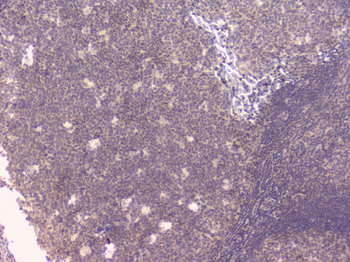
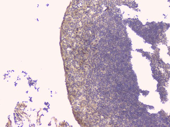
IHC analysis of TNFRSF11B using anti-TNFRSF11B antibody. TNFRSF11B was detected in paraffin-embedded section of human tonsil tissues. Heat mediated antigen retrieval was performed in citrate buffer (pH6, epitope retrieval solution) for 20 mins. The tissue section was blocked with 10% goat serum. The tissue section was then incubated with 1 µg/ml rabbit anti-TNFRSF11B Antibody overnight at 4°C. Biotinylated goat anti-rabbit IgG was used as secondary antibody and incubated for 30 minutes at 37°C. The tissue section was developed using Strepavidin-Biotin-Complex (SABC) with DAB as the chromogen.

IHC analysis of TNFRSF11B using anti-TNFRSF11B antibody. TNFRSF11B was detected in paraffin-embedded section of human tonsil tissues. Heat mediated antigen retrieval was performed in citrate buffer (pH6, epitope retrieval solution) for 20 mins. The tissue section was blocked with 10% goat serum. The tissue section was then incubated with 1 µg/ml rabbit anti-TNFRSF11B Antibody overnight at 4°C. Biotinylated goat anti-rabbit IgG was used as secondary antibody and incubated for 30 minutes at 37°C. The tissue section was developed using Strepavidin-Biotin-Complex (SABC) with DAB as the chromogen.

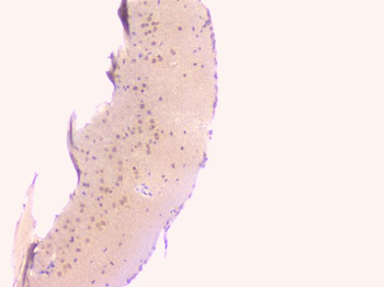
IHC analysis of TNFRSF11B using anti-TNFRSF11B antibody. TNFRSF11B was detected in paraffin-embedded section of mouse brain tissues. Heat mediated antigen retrieval was performed in citrate buffer (pH6, epitope retrieval solution) for 20 mins. The tissue section was blocked with 10% goat serum. The tissue section was then incubated with 1 µg/ml rabbit anti-TNFRSF11B Antibody overnight at 4°C. Biotinylated goat anti-rabbit IgG was used as secondary antibody and incubated for 30 minutes at 37°C. The tissue section was developed using Strepavidin-Biotin-Complex (SABC) with DAB as the chromogen.

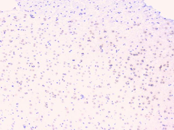
IHC analysis of TNFRSF11B using anti-TNFRSF11B antibody. TNFRSF11B was detected in paraffin-embedded section of rat brain tissues. Heat mediated antigen retrieval was performed in citrate buffer (pH6, epitope retrieval solution) for 20 mins. The tissue section was blocked with 10% goat serum. The tissue section was then incubated with 1 µg/ml rabbit anti-TNFRSF11B Antibody overnight at 4°C. Biotinylated goat anti-rabbit IgG was used as secondary antibody and incubated for 30 minutes at 37°C. The tissue section was developed using Strepavidin-Biotin-Complex (SABC) with DAB as the chromogen.

IHC analysis of TNFRSF11B using anti-TNFRSF11B antibody. TNFRSF11B was detected in paraffin-embedded section of rat brain tissues. Heat mediated antigen retrieval was performed in citrate buffer (pH6, epitope retrieval solution) for 20 mins. The tissue section was blocked with 10% goat serum. The tissue section was then incubated with 1 µg/ml rabbit anti-TNFRSF11B Antibody overnight at 4°C. Biotinylated goat anti-rabbit IgG was used as secondary antibody and incubated for 30 minutes at 37°C. The tissue section was developed using Strepavidin-Biotin-Complex (SABC) with DAB as the chromogen.

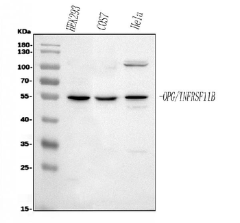
Western blot analysis of TNFRSF11B using anti-TNFRSF11B antibody. Electrophoresis was performed on a 5-20% SDS-PAGE gel at 70V (Stacking gel) / 90V (Resolving gel) for 2-3 hours. The sample well of each lane was loaded with 30 ug of sample under reducing conditions. Lane 1: human HEK293 whole cell lysates, Lane 2: monkey COS-7 whole cell lysates, Lane 3: human Hela whole cell lysates. After electrophoresis, proteins were transferred to a nitrocellulose membrane at 150 mA for 50-90 minutes. Blocked the membrane with 5% non-fat milk/TBS for 1.5 hour at RT. The membrane was incubated with rabbit anti-TNFRSF11B antigen affinity purified polyclonal antibody at 0.5 µg/mL overnight at 4°C, then washed with TBS-0.1% Tween 3 times with 5 minutes each and probed with a goat anti-rabbit IgG-HRP secondary antibody at a dilution of 1:5000 for 1.5 hour at RT. The signal is developed using an Enhanced Chemiluminescent detection (ECL) kit with Tanon 5200 system. A specific band was detected for TNFRSF11B at approximately 55 kDa. The expected band size for TNFRSF11B is at 46 kDa.
Quick Database Links
UniProt Details
−Documents Download
Request a Document
Protocol Information
Osteoprotegerin/TNFRSF11B Rabbit Polyclonal Antibody (orb570415)
Participating in our Biorbyt product reviews program enables you to support fellow scientists by sharing your firsthand experience with our products.
Login to Submit a ReviewFilter by Rating
- 5 stars
- 4 stars
- 3 stars
- 2 stars
- 1 stars
Filter by Applications
Filter by Species
The antibody was used for WB detection of cell lysate samples and showed excellent specificity. Only a single target band was observed on the membrane, with no additional non-specific bands or tailing. The background was very low, eliminating the need for prolonged washing steps. The antibody showed strong sensitivity, producing clear signals even in low-expression samples. The bands were sharp with good grayscale separation, and the data were suitable for direct statistical analysis in publications. The recommended dilution ratio in the datasheet showed good adaptability, and the incubation conditions were flexible, allowing even beginners to obtain consistent results. In three consecutive independent repeat experiments, the band intensity and position remained highly consistent, demonstrating reliable batch-to-batch stability. . The product was securely packaged, shipped under cold-chain conditions, and arrived without leakage. It has been added to the laboratory’s long-term purchasing list.
The sensitivity of this antibody exceeded expectations. It is highly suitable for detecting low-abundance proteins and can generate clear, strong signals even with low sample loading amounts. Previous antibodies often resulted in high background and weak signals, but this antibody effectively resolved these issues, producing clean and reproducible bands. For laboratories studying signaling pathways or working with low-abundance samples, this antibody is a valuable tool and is highly recommended for researchers facing similar challenges.
The WB antibody purchased this time performed exceptionally well. The antibody showed high specificity, producing a single, clear band without non-specific bands or tailing. The background was very clean, with an ideal signal-to-noise ratio, and the observed molecular weight matched the expected size. Batch-to-batch consistency was excellent, and the experimental results were clear and reliable. There was no need to repeatedly question whether the band was correct, greatly improving confidence in the data. This antibody is an essential reagent for WB experiments.