You have no items in your shopping cart.
Featured
Description
Research Area
Immunology & Inflammation
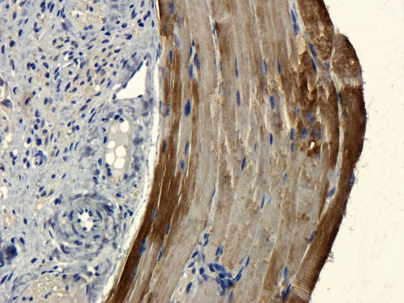
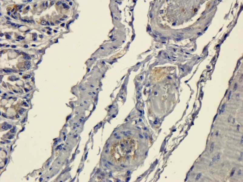
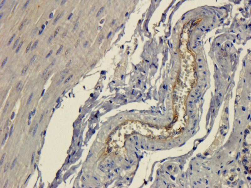
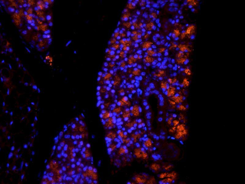
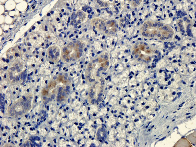
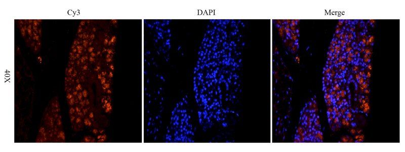
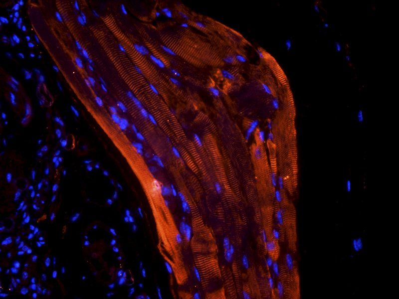
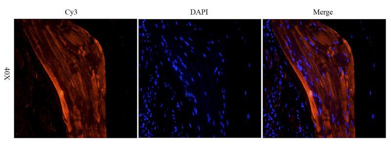
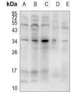
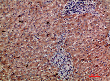
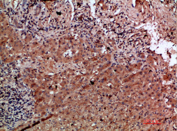
Images & Validation
−


Item 1 of 12
| Tested Applications | ELISA, ICC, IF, IHC-P, WB |
|---|---|
| Dilution Range | WB: 1-2μg/ml, IF/ICC: 1:100, IHC-P: 1:200 (based on 0.5 mg/ml) |
| Reactivity | Human, Mouse, Porcine, Rat |
| Application Notes |
Key Properties
−| Host | Rabbit |
|---|---|
| Clonality | Polyclonal |
| Isotype | IgG |
| Immunogen | KLH conjugated synthetic peptide derived from human CD303. Please contact us for the exact immunogen sequence. The peptide is available as orb374772. |
| Target | CD303 |
| Molecular Weight | 25 kDa |
| Purity | Polyclonal antibodies are purified by peptide affinity chromatography |
| Conjugation | Unconjugated |
Storage & Handling
−| Storage | Maintain refrigerated at 2-8°C for up to 2 weeks. For long term storage store at -20°C in small aliquots to prevent freeze-thaw cycles. |
|---|---|
| Form/Appearance | 10 mM PBS, 0.02% sodium azide |
| Concentration | - 100 μg (in 200 μl): 0.5 mg/ml- 200 μg (in 400 μl): 0.5 mg/ml |
| Expiration Date | 12 months from date of receipt. |
| Disclaimer | For research use only |
Alternative Names
−Anti-BDCA-2 antibody, anti-CLEC4C antibody, anti-BDCA2 antibody, anti-Blood dendritic cell antigen 2 antibody, anti-Blood dendritic cell antigen 2 protein antibody, anti-C-type (calcium dependent antibody, anti-carbohydrate-recognition domain) lectin antibody, anti-superfamily member 11 antibody, anti-C-type (calcium dependent antibody, anti-carbohydrate-recognition domain) lectin antibody, anti-superfamily member 7 antibody, anti-C-type lectin domain family 4 member C antibody, anti-C-type lectin domain family 4 antibody, anti-member C antibody, anti-isoform 1 antibody, anti-C-type lectin superfamily member 7 antibody, anti-CD303 antibody, anti-CD303 antigen antibody, anti-CLECSF11 antibody, anti-CLECSF7 antibody, anti-Dendritic cell lectin antibody, anti-dendritic cell lectin b antibody, anti-Dendritic lectin antibody, anti-DLEC antibody, anti-HECL antibody, anti-Lectin antibody, anti-C-type antibody, anti-superfamily member 11 antibody, anti-MGC125789 antibody, anti-MGC125791 antibody, anti-MGC125792 antibody, anti-MGC125793 antibody, anti-PRO34150 antibody.
Similar Products
−- Item 1 of 1
CD303 Rabbit Polyclonal Antibody [orb666327]
WB
Human, Mouse, Rat
Rabbit
Polyclonal
Unconjugated
100 μl, 200 μl, 50 μl, 30 μl 
CD303 Rabbit Polyclonal Antibody (FITC) [orb397146]
ICC, IF
Human, Mouse, Rat
Rabbit
Polyclonal
FITC
100 μg- Item 1 of 2
CLEC4C Antibody [orb1277329]
IHC
Human
Rabbit
Polyclonal
Unconjugated
Quality Guarantee
Explore bioreagents carefree to elevate your research. All our products are rigorously tested for performance. If a product does not perform as described on its datasheet, our scientific support team will provide expert troubleshooting, a prompt replacement, or a refund. For full details, please see our Terms & Conditions and Buying Guide. Contact us at support@biorbyt.com.
Protocol Information
WB
Western Blot (IB, immunoblot)
IHC-P
Immunohistochemistry Paraffin
IF
Immunofluorescence
ICC
Immunocytochemistry
ELISA
Enzyme-linked Immunosorbent Assay (EIA)
Available Sizes
Select a size below
Choose Conjugation or Carrier Free Version
Free Secondary Antibody (20 ul)0/0
Please add an antibody product to your cart first.