You have no items in your shopping cart.
Description
Research Area
Immunology & Inflammation
Images & Validation
−Item 1 of 5
| Tested Applications | IHC-P, WB |
|---|---|
| Dilution Range | WB - 1:1000, IHC-P - 1:10-50 |
| Reactivity | Human, Mouse |
Key Properties
−| Antibody Type | Primary Antibody |
|---|---|
| Host | Rabbit |
| Clonality | Polyclonal |
| Isotype | Rabbit IgG |
| Immunogen | This CD73 (NT5E) antibody is generated from rabbits immunized with a KLH conjugated synthetic peptide between 520-550 amino acids from the C-terminal region of human CD73 (NT5E). Antigen Region: 520-550 aa. |
| Target | NT5E |
| Molecular Weight | 63368 Da |
| Conjugation | Unconjugated |
Storage & Handling
−| Storage | Maintain refrigerated at 2-8°C for up to 2 weeks. For long term storage store at -20°C in small aliquots to prevent freeze-thaw cycles |
|---|---|
| Form/Appearance | Purified polyclonal antibody supplied in PBS with 0.09% (W/V) sodium azide. This antibody is prepared by Saturated Ammonium Sulfate (SAS) precipitation followed by dialysis against PBS. |
| Expiration Date | 12 months from date of receipt. |
| Disclaimer | For research use only |
Alternative Names
−5'-nucleotidase, 5'-NT, Ecto-5'-nucleotidase, CD73, NT5E, NT5, NTE
Similar Products
−- Item 1 of 5

Quality Guarantee
Explore bioreagents carefree to elevate your research. All our products are rigorously tested for performance. If a product does not perform as described on its datasheet, our scientific support team will provide expert troubleshooting, a prompt replacement, or a refund. For full details, please see our Terms & Conditions and Buying Guide. Contact us at support@biorbyt.com.
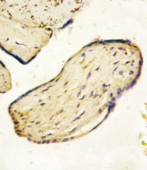
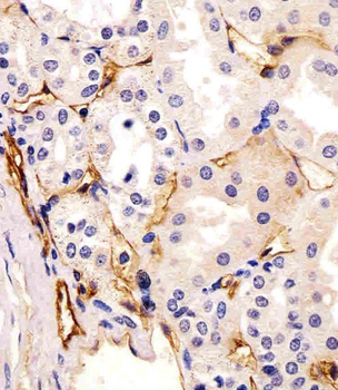
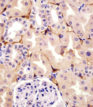
Formalin-fixed and paraffin-embedded human Placenta tissue reacted with CD73 (NT5E) Antibody (C-term), which was peroxidase-conjugated to the secondary antibody, followed by AEC staining. This data demonstrates the use of this antibody for immunohistochemistry; clinical relevance has not been evaluated.
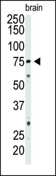
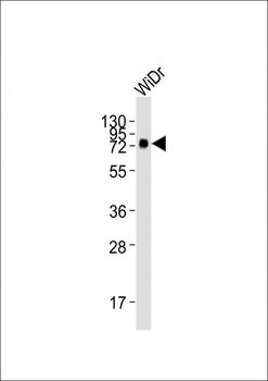
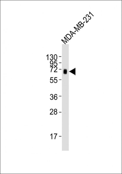
Western blot analysis of CD73 Pab in mouse brain tissue lysate. CD73 (Arrow) was detected using purified Pab. Secondary HRP-anti-rabbit was used for signal visualization with chemiluminescence.
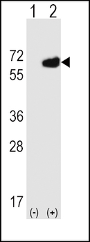
Western blot analysis of NT5E (arrow) using rabbit polyclonal NT5E Antibody (M535). 293 cell lysates (2 ug/lane) either nontransfected (Lane 1) or transiently transfected (Lane 2) with the NT5E gene.
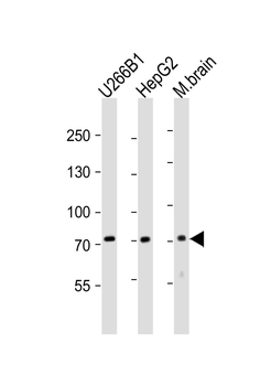
Western blot analysis of lysates from U266B1, HepG2 cell line, mouse brain tissue lysate (from left to right), using NT5E-M535 Antibody. Diluted at 1:2000 at each lane. A goat anti-rabbit IgG H&L (HRP) at 1:10000 dilution was used as the secondary antibody. Lysates at 20 ug per lane.
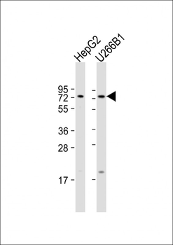
All lanes: Anti-NT5E Antibody (M535) at 1:1000 dilution. Lane 1: HepG2 whole cell lysate. Lane 2: U266B1 whole cell lysate. Lysates/proteins at 20 µg per lane. Secondary Goat Anti-Rabbit IgG, (H+L), Peroxidase conjugated at 1/10000 dilution. Predicted band size: 63 kDa. Blocking/Dilution buffer: 5% NFDM/TBST.
Quick Database Links
UniProt Details
− No UniProt data available
NCBI Reference Sequences
−Associated Accession Numbers
Curated reference sequences for the gene transcript and protein product| Protein | NP_002517.1, NP_001191742.1 |
|---|
Documents Download
Datasheet
Product Information
Request a Document
Protocol Information
WB
Western Blot (IB, immunoblot)
IHC-P
Immunohistochemistry Paraffin
CD73 (NT5E) Antibody (C-term) (orb1931872)

- 0.0
Based on 0 reviews
Participating in our Biorbyt product reviews program enables you to support fellow scientists by sharing your firsthand experience with our products.
Login to Submit a ReviewAvailable Sizes
Select a size below
Choose Conjugation or Carrier Free Version
Free Secondary Antibody (20 ul)0/0
Please add an antibody product to your cart first.