You have no items in your shopping cart.
Description
Research Area
Epigenetics
Images & Validation
−Item 1 of 6
| Tested Applications | ELISA, IF, IHC-P, WB |
|---|---|
| Dilution Range | WB: 1:500-2000, IHC-P: 1:50-200, IF/ICC: 1:20-100 |
| Reactivity | Human, Mouse, Rat |
Key Properties
−| Host | Rabbit |
|---|---|
| Clonality | Polyclonal |
| Isotype | IgG |
| Immunogen | A synthetic peptide corresponding to a sequence within amino acids 1-164 of human CDKN1A/p21CIP1 (NP_000380.1). |
| Molecular Weight | 18kDa |
| Purification | Affinity purification |
| Conjugation | Unconjugated |
Storage & Handling
−| Storage | Maintain refrigerated at 2-8°C for up to 2 weeks. For long term storage store at -20°C in small aliquots to prevent freeze-thaw cycles. |
|---|---|
| Buffer/Preservatives | PBS with 0.09% Sodium azide,50% glycerol,pH7.3. |
| Concentration | 1 mg/ml |
| Expiration Date | 12 months from date of receipt. |
| Disclaimer | For research use only |
Alternative Names
−anti CAP20 antibody, anti CDKN1 antibody, anti CIP1 antibody, anti MDA-6 antibody, anti P21 antibody, anti SDI1 antibody, anti WAF1 antibody, anti p21CIP1 antibody
Quality Guarantee
Explore bioreagents carefree to elevate your research. All our products are rigorously tested for performance. If a product does not perform as described on its datasheet, our scientific support team will provide expert troubleshooting, a prompt replacement, or a refund. For full details, please see our Terms & Conditions and Buying Guide. Contact us at support@biorbyt.com.
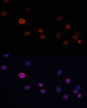
Immunofluorescence analysis of C6 cells using CDKN1A/p21CIP1 Rabbit pAb (orb48324) at dilution of 1:100. Blue: DAPI for nuclear staining.
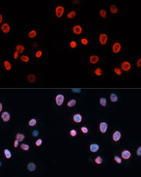
Immunofluorescence analysis of HeLa cells using CDKN1A/p21CIP1 Rabbit pAb (orb48324) at dilution of 1:100. Blue: DAPI for nuclear staining.
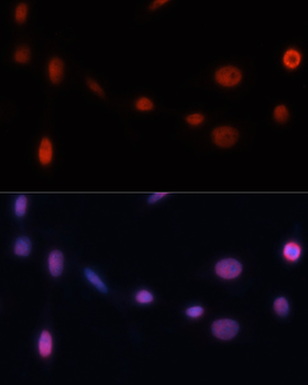
Immunofluorescence analysis of NIH/3T3 cells using CDKN1A/p21CIP1 Rabbit pAb (orb48324) at dilution of 1:100. Blue: DAPI for nuclear staining.
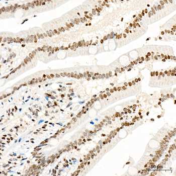
Immunohistochemistry analysis of paraffin-embedded Human colon tissue using CDKN1A/p21CIP1 Rabbit pAb (orb48324) at a dilution of 1:200 (40x lens). High pressure antigen retrieval performed with 0.01M Citrate Bufferr (pH 6.0) prior to IHC staining.
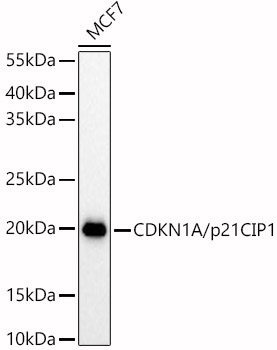
Western blot analysis of lysates from MCF7 cells, using CDKN1A/p21CIP1 Rabbit pAb (orb48324) at 1:900 dilution. Secondary antibody: HRP-conjugated Goat anti-Rabbit IgG (H+L) at 1:10000 dilution. Lysates/proteins: 25 µg per lane. Blocking buffer: 3% nonfat dry milk in TBST. Detection: ECL Basic Kit. Exposure time: 180s.
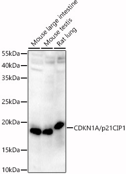
Western blot analysis of various lysates, using CDKN1A/p21CIP1 Rabbit pAb (orb48324) at 1:900 dilution. Secondary antibody: HRP-conjugated Goat anti-Rabbit IgG (H+L) at 1:10000 dilution. Lysates/proteins: 25 µg per lane. Blocking buffer: 3% nonfat dry milk in TBST. Detection: ECL Basic Kit. Exposure time: 180s.
Quick Database Links
UniProt
UniProt Details
− No UniProt data available
Documents Download
Datasheet
Product Information
Request a Document
Protocol Information
WB
Western Blot (IB, immunoblot)
IHC-P
Immunohistochemistry Paraffin
IF
Immunofluorescence
ELISA
Enzyme-linked Immunosorbent Assay (EIA)
Filter by Applications
Filter by Species
Kimoto, Saki et al. Nacre extract derived from Pinctada fucata mitigates skeletal muscle aging by suppressing senescence-associated inflammation and preserving contractile features in naturally aged mice and C2C12 myotubes Asian Pacific Journal of Tropical Biomedicine, (2026)
Applications
IF
Reactivity
Mouse
CDKN1A/p21CIP1 Rabbit pAb (orb48324)

- 0.0
Based on 0 reviews
Participating in our Biorbyt product reviews program enables you to support fellow scientists by sharing your firsthand experience with our products.
Login to Submit a Review