You have no items in your shopping cart.
CFL1 Antibody (Center)
SKU: orb1931349
Description
Research Area
Cancer Biology, Cardiovascular Research, Immunology & Inflammation, Neuroscience, Signal Transduction
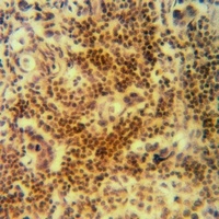
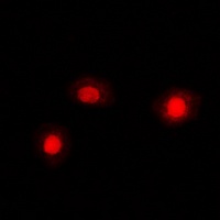
Images & Validation
−Item 1 of 5
| Tested Applications | FC, WB |
|---|---|
| Dilution Range | WB - 1:1000, FC - 1:10-50 |
| Reactivity | Human, Mouse |
| Predicted Reactivity | Bovine, Gallus, Monkey, Porcine, Rat, Xenopus |
Key Properties
−| Antibody Type | Primary Antibody |
|---|---|
| Host | Rabbit |
| Clonality | Polyclonal |
| Isotype | Rabbit IgG |
| Molecular Weight | 18502 Da |
| Conjugation | Unconjugated |
Storage & Handling
−| Storage | Maintain refrigerated at 2-8°C for up to 2 weeks. For long term storage store at -20°C in small aliquots to prevent freeze-thaw cycles |
|---|---|
| Form/Appearance | Purified polyclonal antibody supplied in PBS with 0.09% (W/V) sodium azide. This antibody is purified through a protein A column, followed by peptide affinity purification. |
| Expiration Date | 12 months from date of receipt. |
| Disclaimer | For research use only |
Alternative Names
−CFL
Similar Products
−- Item 1 of 3
Cofilin Rabbit Polyclonal Antibody [orb393241]
IF, IHC, WB
Human, Mouse, Porcine, Rat, Sheep, Zebrafish
Rabbit
Polyclonal
Unconjugated
30 μl, 100 μl, 200 μl, 50 μl
Quality Guarantee
Explore bioreagents carefree to elevate your research. All our products are rigorously tested for performance. If a product does not perform as described on its datasheet, our scientific support team will provide expert troubleshooting, a prompt replacement, or a refund. For full details, please see our Terms & Conditions and Buying Guide. Contact us at support@biorbyt.com.
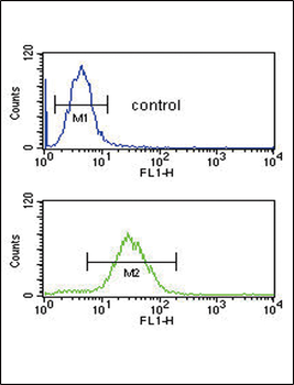
CFL1 Antibody (Center) flow cytometric analysis of HL-60 cells (bottom histogram) compared to a negative control cell (top histogram). FITC-conjugated goat-anti-rabbit secondary antibodies were used for the analysis.
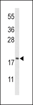
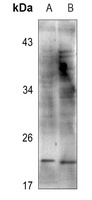
Western blot analysis of CFL1 Antibody (Center) in HL-60 cell line lysates (35 ug/lane). CFL1 (arrow) was detected using the purified Pab;
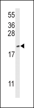
Western blot analysis of CFL1 Antibody (Center) in mouse NIH-3T3 cell line lysates (35 ug/lane). CFL1 (arrow) was detected using the purified Pab.
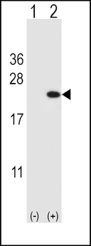
Western blot analysis of CFL1 (arrow) using rabbit polyclonal CFL1 Antibody (Center). 293 cell lysates (2 ug/lane) either nontransfected (Lane 1) or transiently transfected (Lane 2) with the CFL1 gene.
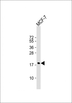
Anti-CFL1 Antibody (Center) at 1:1000 dilution + MCF-7 whole cell lysate. Lysates/proteins at 20 µg per lane. Secondary Goat Anti-Rabbit IgG, (H+L), Peroxidase conjugated at 1/10000 dilution. Predicted band size: 19 kDa. Blocking/Dilution buffer: 5% NFDM/TBST.
Quick Database Links
UniProt
UniProt Details
− No UniProt data available
Documents Download
Datasheet
Product Information
Request a Document
Protocol Information
WB
Western Blot (IB, immunoblot)
FC
Flow Cytometry
CFL1 Antibody (Center) (orb1931349)

- 0.0
Based on 0 reviews
Participating in our Biorbyt product reviews program enables you to support fellow scientists by sharing your firsthand experience with our products.
Login to Submit a ReviewAvailable Sizes
Select a size below