You have no items in your shopping cart.
KO/KD
Validated
Validated
Description
Images & Validation
−Item 1 of 4
| Tested Applications | HAI, KO/KD Validated |
|---|---|
| Application Notes |
Key Properties
−| Antibody Type | Primary Antibody |
|---|---|
| Host | Rabbit |
| Clonality | Polyclonal |
| Isotype | Antiserum |
| Immunogen | Chicken washed pooled Red Blood Cells (RBC) |
| Purity | This product was prepared from polyspecific antiserum by a delipidation and defibrination. |
| Conjugation | Unconjugated |
Storage & Handling
−| Storage | Store vial at 4° C prior to restoration. For extended storage aliquot contents and freeze at -20° C or below. Avoid cycles of freezing and thawing. Centrifuge product if not completely clear after standing at room temperature. This product is stable for several weeks at 4° C as an undiluted liquid. Dilute only prior to immediate use. |
|---|---|
| Form/Appearance | Lyophilized |
| Buffer/Preservatives | 0.01% (w/v) Sodium Azide |
| Concentration | 60 mg/mL |
| Expiration Date | 12 months from date of receipt. |
| Disclaimer | For research use only |
Alternative Names
−Anti-RBC antibody, Red Blood Cell Antibody, Antibody for hemagglutination, rabbit anti RBC, rabbit anti-chicken Red Blood Cells (RBC), haemolysin, hemolysin, erythrocytes sensitizing agent
Similar Products
−Quality Guarantee
Explore bioreagents carefree to elevate your research. All our products are rigorously tested for performance. If a product does not perform as described on its datasheet, our scientific support team will provide expert troubleshooting, a prompt replacement, or a refund. For full details, please see our Terms & Conditions and Buying Guide. Contact us at support@biorbyt.com.
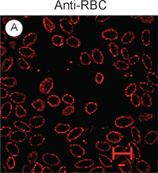
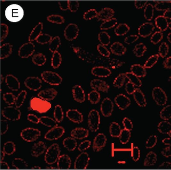
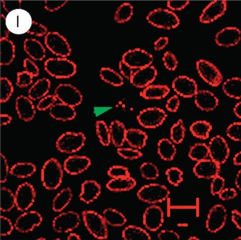
Confocal images of infected chicken blood cells stained with anti-red blood cell antibodies. Anti-RBC, images stained with rabbit anti-chicken red blood cell antibody 103–4139; DIC, differential interference contrast images; Hoechst, Hoechst dye staining of nuclei (DNA); Merged, merged images of anti-RBC, DIC, and DAPI. (A-H) Two cells infected with L. sabrazesi gametocytes have some red dots (yellow arrowheads) that appear to be within the cytoplasm of the host cells. (E-H) A strongly stained white cell (grey arrowheads) that has two nuclei and rough granules in the cytoplasm, suggesting heterophils, monocytes, macrophages, or eosinophils. (I-L) A small cell that has a round nucleus and red dots similar (green arrowheads) to those seen in the infected cells. The small size of the cell suggests that it is likely a thrombocyte. The red ruler in each image indicates 10 µm.
Confocal images of infected chicken blood cells stained with anti-red blood cell antibodies.Anti-RBC, images stained with rabbit anti-chicken red blood cell antibody 103–4139; DIC, differential interference contrast images; Hoechst, Hoechst dye staining of nuclei (DNA); Merged, merged images of anti-RBC, DIC, and DAPI. (A-H) Two cells infected with L. sabrazesi gametocytes have some red dots (yellow arrowheads) that appear to be within the cytoplasm of the host cells. (E-H) A strongly stained white cell (grey arrowheads) that has two nuclei and rough granules in the cytoplasm, suggesting heterophils, monocytes, macrophages, or eosinophils. (I-L) A small cell that has a round nucleus and red dots similar (green arrowheads) to those seen in the infected cells. The small size of the cell suggests that it is likely a thrombocyte. The red ruler in each image indicates 10 µm.
Confocal images of infected chicken blood cells stained with anti-red blood cell antibodies.Anti-RBC, images stained with rabbit anti-chicken red blood cell antibody 103–4139; DIC, differential interference contrast images; Hoechst, Hoechst dye staining of nuclei (DNA); Merged, merged images of anti-RBC, DIC, and DAPI. (A-H) Two cells infected with L. sabrazesi gametocytes have some red dots (yellow arrowheads) that appear to be within the cytoplasm of the host cells. (E-H) A strongly stained white cell (grey arrowheads) that has two nuclei and rough granules in the cytoplasm, suggesting heterophils, monocytes, macrophages, or eosinophils. (I-L) A small cell that has a round nucleus and red dots similar (green arrowheads) to those seen in the infected cells. The small size of the cell suggests that it is likely a thrombocyte. The red ruler in each image indicates 10 µm.
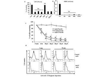
Therapeutic efficacy of anti-human P mAb 19.1 in a murine model of extravascular hemolysisa: Pharmacodynamics of mAb 19.1 in hP transgenic mice. Each mouse was treated with 0.5 mg mAb 19.1 (n = 3 mice). Serum samples were collected before and at various time points after mAb treatment and assessed for LPS-dependent AP complement activity. At this dosage of mAb 19.1, AP complement activity was suppressed to background (P−/−) level for 2 days. EDTA: time 0 sample with EDTA (20 mM) added. NS, non-significant comparing 8, 24 and 48 hr samples with fP−/− or EDTA-treated serum, or comparing 72 hr sample with 0 hr sample. One-way ANOVA. b: Pharmacodynamics of anti-mouse C5 mAb (BB5.1) in hP transgenic mice. Each mouse was treated with 1mg of anti-mouse C5 mAb (n = 3). Serum samples were collected before and at various time points after mAb treatment and assessed for lytic activity using antibody-sensitized chicken RBCs. C5 knockout (KO) mouse serum was used as a control for C5 inhibition. Percentage of chicken RBC (cRBC) lysis was normalized to a sample completely lysed by hypotonic shock in double distilled water. * P < 0.0001, NS: non-significant compared with C5KO serum. One-way ANOVA. c. Effect of anti-hP mAb 19.1 on the survival of transfused CFSC-labeled DAF−/−/Crry−/−/C3−/− mouse erythrocytes in hP- transgenic mice. Recipient mice were treated with anti-hP mAb 19.1 (n = 4) or an isotype control mAb (n = 4) or anti-mouse C5 mAb (n = 3) 6 hours prior to red blood cell transfusion and blood samples were taken at 5 min, 6 hrs and then daily for 5 days. The percentage of CFSC-labeled red blood cells was measured by FACS and normalized to that determined at 5 min (100%). Transfused DAF−/−/Crry−/−/C3−/− mouse red blood cells were rapidly eliminated in control mAb- or anti-C5 mAb-treated hP transgenic mice but such an outcome was prevented by mAb 19.1 treatment. * P < 0.001, Two-way ANOVA. d. FACS analysis of activated C3 fragment deposition on DAF−/−/Crry−/−/C3−/− RBCs 5 and 30 min after their transfusion into control mAb- or mAb 19.1-treated hP-Tg/P−/− mice (representative data from two recipient mice are shown). At both time points, C3 fragment deposition was significantly higher on RBCs transfused into control mAb-treated than mAb 19.1-treated hP-Tg/P−/− mice. The reason for the marked reduction in C3 fragment deposition on RBCs in control mAb-treated hP-Tg/P−/− mice between 5 and 30 min is unknown, but could be caused by C3 fragment degradation to C3d or shedding from the cell surface or rapid removal of the C3-opsonized cells. Data in a-c are presented as mean (SD) with indicated n numbers. Antibody-sensitized chicken RBC (p/n orb1567092) prepared by incubating the cells with a rabbit anti-chicken RBC antibody (p/n orb1568264).
Documents Download
Datasheet
Product Information
Request a Document
Chicken Red Blood Cell RBC Antibody (orb1568264)

- 0.0
Based on 0 reviews
Participating in our Biorbyt product reviews program enables you to support fellow scientists by sharing your firsthand experience with our products.
Login to Submit a ReviewAvailable Sizes
Select a size below
Free Secondary Antibody (20 ul)0/0
Please add an antibody product to your cart first.