You have no items in your shopping cart.
Description
Research Area
Musculoskeletal & Connective Tissue Research
Images & Validation
−Item 1 of 3
| Tested Applications | ELISA, FC, IHC, IP, WB |
|---|---|
| Dilution Range | ELISA: 1:3,000 - 1:6,000, FC: 5µg/mL, IHC: 1:50 - 1:200, IP: 1:100, WB: 1:3,000 - 1:6,000 |
| Reactivity | Bovine, Human, Mouse, Rat |
| Application Notes |
Key Properties
−| Antibody Type | Primary Antibody |
|---|---|
| Host | Rabbit |
| Clonality | Polyclonal |
| Isotype | IgG |
| Immunogen | Collagen Type I from human and bovine placenta. |
| Target | COL1A1 |
| Purity | This product has been prepared by immunoaffinity chromatography using immobilized antigens. Some class-specific anti-collagens may be specific for three-dimensional epitopes which may result in diminished reactivity with denatured collagen or formalin-fixed, paraffin embedded tissues. This antibody reacts with most mammalian Type I collagens and has expected cross-reactivity with Type III and negligible cross reactivity with Type II, IV, V or VI collagens. Non-specific cross-reaction of anti-collagen antibodies with other human serum proteins or non-collagen extracellular matrix proteins has not been tested. |
| Conjugation | FITC |
Storage & Handling
−| Storage | Store vial at 4° C prior to restoration. Restore with 0.05 mL of deionized water (or equivalent). For extended storage aliquot contents and freeze at -20° C or below. Avoid cycles of freezing and thawing. Centrifuge product if not completely clear after standing at room temperature. This product is stable for several weeks at 4° C as an undiluted liquid. Dilute only prior to immediate use. Expiration date is one (1) year from date of restoration. |
|---|---|
| Form/Appearance | Lyophilized |
| Buffer/Preservatives | Preservative: 0.01% (w/v) Sodium Azide. Stabilizer: 10 mg/mL Bovine Serum Albumin (rAlbumin) - Immunoglobulin and Protease free; Buffer: 0.01 M Sodium Phosphate, 0.25 M Sodium Chloride, pH 7.2 |
| Concentration | 1.0 mg/mL |
| Expiration Date | 12 months from date of receipt. |
| Disclaimer | For research use only |
Alternative Names
−rabbit anti-collagen type I antibody fluorescein conjugation, FITC conjugated rabbit anti-collagen type I antibody, Collagen Of Skin Tendon And Bone, Collagen Type 1 antibody, Collagen type I alpha 1 antibody, Collagen alpha-1 (I) chain, Alpha-1 type I collagen, type 1 procollagen alpha 1
Similar Products
−- Item 1 of 1
Collagen I Rabbit Polyclonal Antibody (FITC) [orb15412]
ICC, IF
Human, Mouse, Rat
Rabbit
Polyclonal
FITC
100 μg 
Collagen I/COL1A1 Rabbit Polyclonal Antibody (FITC) [orb2575100]
Human, Mouse, Rat
Rabbit
Polyclonal
FITC
100 μg
Quality Guarantee
Explore bioreagents carefree to elevate your research. All our products are rigorously tested for performance. If a product does not perform as described on its datasheet, our scientific support team will provide expert troubleshooting, a prompt replacement, or a refund. For full details, please see our Terms & Conditions and Buying Guide. Contact us at support@biorbyt.com.
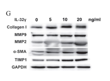
Effect of different concentrations (0, 5, 10 and 20 ng/mL) of IL‑32γ on LX‑2 activation phenotypes. (G) Western blot analysis was used to measure collagen I, MMP9, MMP2, α‑SMA, TIMP1 and GAPDH expression in whole‑cell extracts.
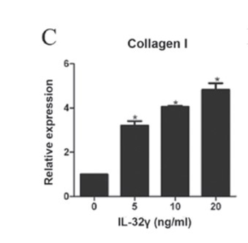
Effect of different concentrations (0, 5, 10 and 20 ng/mL) of IL‑32γ on LX‑2 activation phenotypes. Reverse transcription‑quantitative polymerase chain reaction assessing mRNA levels of α-SMA, (C) collagen I, representing the activation level of LX‑2.
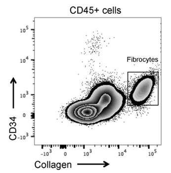
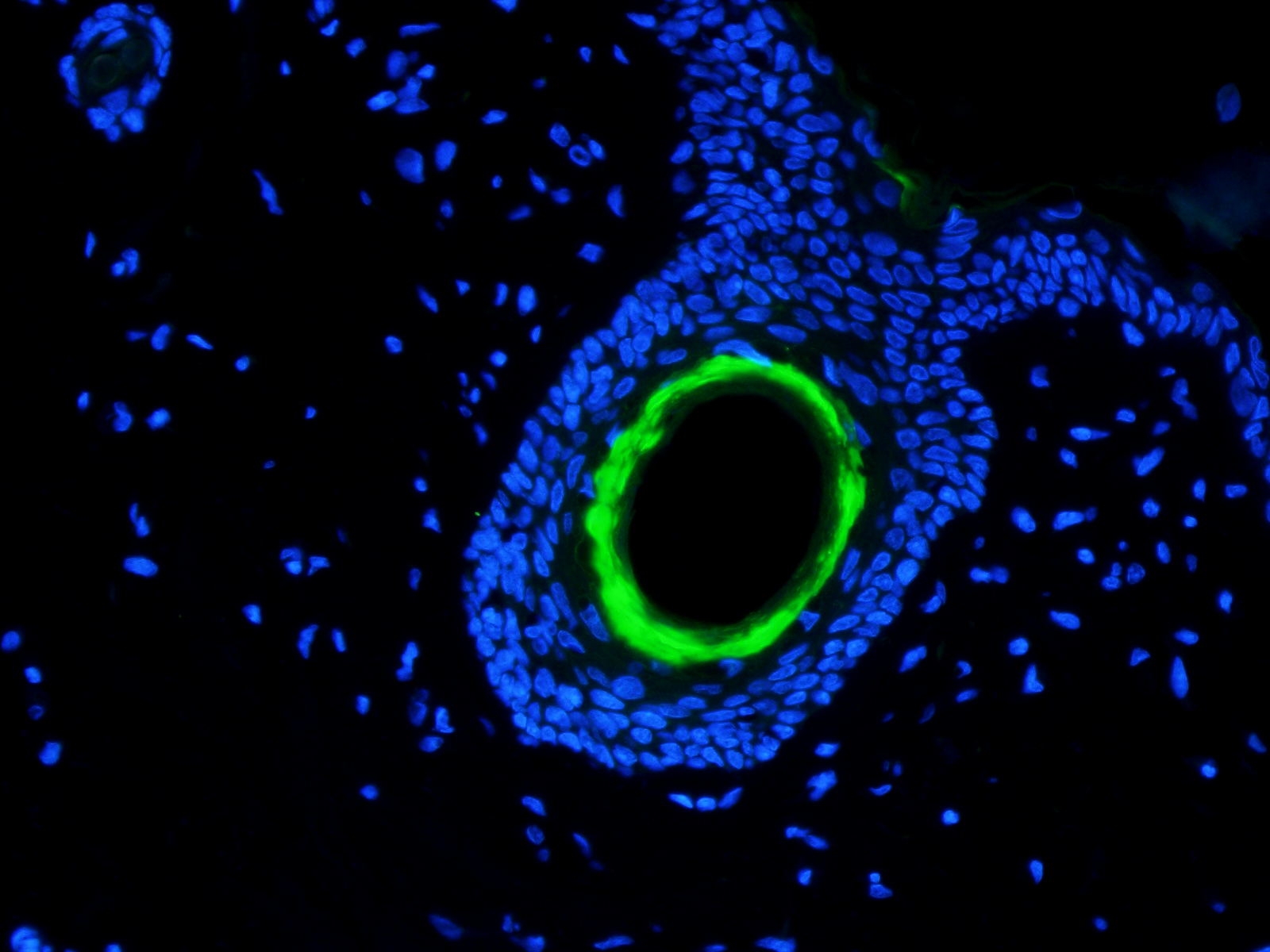
Gating strategy for flow cytometric analysis and cell sorting of circulating fibrocytes using the LIVE/DEAD Fixable Blue Dead Cell Stain kit. For intracellular staining of type I collagen–FITC on B cells cells were fixed and permeabilized with the BD Cytofix/Cytoperm kit.
Quick Database Links
UniProt Details
− No UniProt data available
NCBI Reference Sequences
−Associated Accession Numbers
Curated reference sequences for the gene transcript and protein product| Protein | NP_000079.2 |
|---|
Documents Download
Datasheet
Product Information
Request a Document
Protocol Information
WB
Western Blot (IB, immunoblot)
IHC
Immunohistochemistry
FC
Flow Cytometry
ELISA
Enzyme-linked Immunosorbent Assay (EIA)
IP
Immunoprecipitation
COL1A1 Antibody (FITC) (orb345831)

- 0.0
Based on 0 reviews
Participating in our Biorbyt product reviews program enables you to support fellow scientists by sharing your firsthand experience with our products.
Login to Submit a ReviewAvailable Sizes
Select a size below
Free Secondary Antibody (20 ul)0/0
Please add an antibody product to your cart first.