Cart summary

You have no items in your shopping cart.

CX3CR1 Antibody

SKU: orb1239314
FeaturedFeatured Product

Description

CX3CR1 Antibody

Images & Validation

Tested ApplicationsELISA, FC, IF, IHC-P, WB
ReactivityHuman, Mouse, Rat
Predicted ReactivityBovine

Key Properties

Antibody TypePrimary Antibody
HostRabbit
ClonalityPolyclonal
IsotypeIgG
ImmunogenAnti-CX3CR1 antibody (orb1239314) was raised against a peptide corresponding to 20 amino acids near the amino terminus of mature human CX3CR1. The immunogen is located within the first 50 amino acids of CX3CR1.
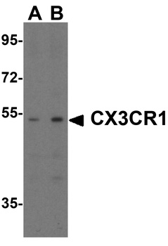
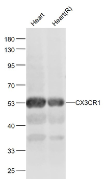
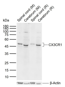
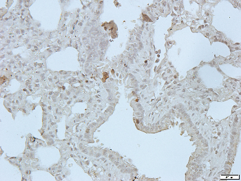
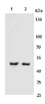
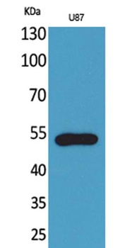
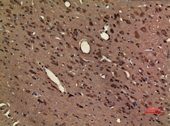
TargetCX3CR1
Molecular WeightPredicted: 40-44 kDObserved: 50 kD
PurificationCX3CR1 Antibody is affinity chromatography purified via peptide column.
ConjugationUnconjugated

Storage & Handling

StorageMaintain refrigerated at 2-8°C for up to 2 weeks. For long term storage store at -20°C in small aliquots to prevent freeze-thaw cycles.
Form/AppearanceLiquid
Buffer/PreservativesCX3CR1 Antibody is supplied in PBS containing 0.02% sodium azide.
Concentration1 mg/mL
DisclaimerFor research use only

Alternative Names

CX3CR1 Antibody: V28, CCRL1, GPR13, CMKDR1, GPRV28, CMKBRL1, CX3C chemokine receptor 1, Beta chemokine receptor-like 1, C-X3-C CKR-1

Similar Products

  • CX3CR1 Rabbit Polyclonal Antibody [orb10490]

    FC,  IF,  IHC-Fr,  IHC-P,  WB

    Bovine, Canine, Human, Rabbit

    Mouse, Rat

    Rabbit

    Polyclonal

    Unconjugated

    50 μl, 100 μl, 200 μl
  • Human Chemokine C-X3-C-Motif Receptor 1 (CX3CR1) ELISA Kit [orb778409]

    Human

    0.16-10 ng/mL

    0.052 ng/mL

    96 T, 48 T
  • Mouse Chemokine C-X3-C-Motif Receptor 1 (CX3CR1) ELISA Kit [orb777096]

    Mouse

    0.16-10 ng/mL

    0.055 ng/mL

    96 T, 48 T
  • Fractalkine Receptor rabbit pAb [orb766717]

    ELISA,  IHC-P,  WB

    Human, Mouse, Rat

    Polyclonal

    Unconjugated

    100 μl, 50 μl
  • CX3CR1 Rabbit Polyclonal Antibody (PE) [orb485433]

    FC,  IF

    Bovine, Canine, Human, Rabbit

    Mouse, Rat

    Rabbit

    Polyclonal

    PE

    100 μl
Quality Guarantee

Quality Guarantee

Explore bioreagents carefree to elevate your research. All our products are rigorously tested for performance. If a product does not perform as described on its datasheet, our scientific support team will provide expert troubleshooting, a prompt replacement, or a refund. For full details, please see our Terms & Conditions and Buying Guide. Contact us at support@biorbyt.com.

CX3CR1 Antibody

Western Blot Validation in Human Cells. Loading: 15 µg of lysates per lane. Antibodies: CX3CR1 orb1239314 (0.5 µg/mL), 1h incubation at RT in 5% NFDM/TBST. Secondary: Goat anti-rabbit IgG HRP conjugate at 1:10000 dilution.

CX3CR1 Antibody

KD Validation in 293 Cells. Loading: 15 µg of lysates per lane. Antibodies: CX3CR1 orb1239314 (0.5 µg/mL), 1h incubation at RT in 5% NFDM/TBST. Secondary: Goat anti-rabbit IgG HRP conjugate at 1:10000 dilution. Lane 1: 293 cells transfected with control siRNAs. Lane 2: 293 cells transfected with CX3CR1 siRNAs.

CX3CR1 Antibody

Western Blot Validation in THP1 Cells. Loading: 15 µg of lysates per lane. Antibodies: CX3CR1 orb1239314 (1 µg/mL), 1h incubation at RT in 5% NFDM/TBST. Secondary: Goat anti-rabbit IgG HRP conjugate at 1:10000 dilution. Lane 1: 0.2 µg/mL, Lane 1: 0.5 µg/mL, Lane 1: 1 µg/mL.

CX3CR1 Antibody

Western Blot Validation in Human Spleen Lysates. Loading: 15 µg of lysates per lane. Antibodies: CX3CR1 orb1239314 (1 µg/mL) in the absence (lane 1) or presence of blocking peptide (lane 2), 1h incubation at RT in 5% NFDM/TBST. Secondary: Goat anti-rabbit IgG HRP conjugate at 1:10000 dilution.

CX3CR1 Antibody

Western Blot Validation in Rat Spleen Tissue. Loading: 15 µg of lysates per lane. Antibodies: CX3CR1 orb1239314, (A; 1 µg/mL, B; 2 µg/mL), 1h incubation at RT in 5% NFDM/TBST. Secondary: Goat anti-rabbit IgG HRP conjugate at 1:10000 dilution.

CX3CR1 Antibody

Flow Cytometry Validation of CX3CR1 in THP-1 Cells. Overlay histogram showing THP-1 cells stained with orb1239314 (red line, 1µg/1x10 6 cells). 1 h incubation at 4°C in 2% FBS/PBS. Followed by secondary antibody 488 goat anti-rabbit IgG (H+L) at 1/500 dilution for 1 h 4°C. Isotype control antibody (Green line) was rabbit IgG1 (1µg/1x10 6 cells) used under the same conditions. Acquisition of > 10000 events was performed.

CX3CR1 Antibody

Immunofluorescence Validation of CX3CR1 In K562 Cells. Immunofluorescent analysis of 4% paraformaldehyde-fixed K562 cells labeling CX3CR1 with orb1239314 at 10 µg/mL, followed by goat anti-rabbit IgG secondary antibody at 1/500 dilution (red) and DAPI staining (blue). Image showing membrane staining on K562 cells.

CX3CR1 Antibody

Immunofluorescence Validation of CX3CR1 in Human Heart. Immunofluorescent analysis of 4% paraformaldehyde-fixed human heart tissue labeling CX3CR1 with orb1239314 at 10 µg/mL, followed by goat anti-rabbit IgG secondary antibody at 1/500 dilution (red).

CX3CR1 Antibody

Immunofluorescence Validation of CX3CR1 in Mouse Heart Tissue. Immunofluorescent analysis of 4% paraformaldehyde-fixed mouse heart tissue labeling CX3CR1 with orb1239314 at 20 µg/mL, followed by goat anti-rabbit IgG secondary antibody at 1/500 dilution (green) and DAPI staining (blue).

CX3CR1 Antibody

Immunohistochemistry Validation of CX3CR1 in Rat Heart Tissue. Immunohistochemical analysis of paraffin-embedded rat heart tissue using anti-CX3CR1 antibody (orb1239314) at 2 µg/ml. Tissue was fixed with formaldehyde and blocked with 10% serum for 1 h at RT; antigen retrieval was by heat mediation with a citrate buffer (pH6). Samples were incubated with primary antibody overnight at 4°C. A goat anti-rabbit IgG H&L (HRP) at 1/250 was used as secondary. Counter stained with Hematoxylin.

CX3CR1 Antibody

Immunofluorescence Validation of CX3CR1 in Rat Heart Tissue. Immunofluorescent analysis of 4% paraformaldehyde-fixed rat heart tissue labeling CX3CR1 with orb1239314 at 20 µg/mL, followed by goat anti-rabbit IgG secondary antibody at 1/500 dilution (green) and DAPI staining (blue).

CX3CR1 Antibody

Immunofluorescence Validation of CX3CR1 in Human Atherosclerosis (Lucas et al., 2003). CX3CR1-positive cells in coronary artery sections was detected by anti-CX3CR1 antibodies by double staining with an anti–smooth muscle actin antibody (yellow).

CX3CR1 Antibody

Regulated Expression Validation of CX3CR1 in Mouse Bone Marrow-derived NK Cells (Barlic et al., 2003). Western blot analysis of CX3CR1 expression detected by anti-CX3CR1 antibodies was induced by IL-2 but was inhibited by IL-15. Mouse skeletal muscle was for negative control and mouse brain was for positive control.

CX3CR1 Antibody

Flow Cytometry Validation of CX3CR1 in Peripheral Blood Mononuclear Cells (PBMCs) of Patients (Yajima et al., 2005). Flow cytometry analysis of CX3CR1 expression detected by anti-CX3CR1 antibodies. Protein expression was more pronounced on CD4 + and CD8 + T cells from patients with untreated active systemic lupus erythematosus (SLE) as compared to those with rheumatoid arthritis (RA) or healthy controls.

CX3CR1 Antibody

CX3CR1 Deficiency Validation in Transgenic Mice (Kumar et al., 2013). Transgenic CX3CR1 gfp (C57BL6/J background) mice, in which either one (CX3CR1 gfp/+) or both (CX3CR1 gfp/gfp) copies of the CX3CR1 gene were interrupted by fluorescent protein (GFP). More CX3CR1 positive cells (green) are found in vascular wall of CX3CR1 gfp/+ mice, but not in CX3CR1 gfp/gfp mice, where cells remained in perivascular region due to leaky microvessels. CX3CR1 expression was detected by anti-CX3CR1 antibodies.

UniProt Details

No UniProt data available

NCBI Reference Sequences

Associated Accession Numbers
Curated reference sequences for the gene transcript and protein product
ProteinNP_001328

Documents Download

Datasheet
Product Information
Download

Request a Document

Protocol Information

WB
Western Blot (IB, immunoblot)
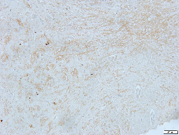
View Protocol
IHC-P
Immunohistochemistry Paraffin
View Protocol
FC
Flow Cytometry
View Protocol
IF
Immunofluorescence
View Protocol
ELISA
Enzyme-linked Immunosorbent Assay (EIA)
View Protocol

CX3CR1 Antibody (orb1239314)

  • Star
  • Star
  • Star
  • Star
  • Star
  • 0.0
Based on 0 reviews

Participating in our Biorbyt product reviews program enables you to support fellow scientists by sharing your firsthand experience with our products.

Login to Submit a Review

No reviews yet

Available Sizes

Select a size below

0.02 mg
$ 290.00
0.1 mg
$ 670.00
DispatchUsually dispatched within 5-10 working days
Bulk Enquiry