You have no items in your shopping cart.
Description
Research Area
Cell Biology
Images & Validation
−Item 1 of 6
| Tested Applications | FC, ICC, IF, IHC, WB |
|---|---|
| Dilution Range | WB (1:1000), IHC (1:100), ICC/IF (1:100), FCM (1:100); optimal dilutions for assays should be determined by the user. |
| Reactivity | Bovine, Drosophila, Frog, Gallus, Human, Mouse, Rat, Sheep |
| Application Notes |
Key Properties
−| Host | Rabbit |
|---|---|
| Clonality | Polyclonal |
| Immunogen | A 35 residue synthetic peptide, corresponding to Rat Erk1 MAP kinase with the CGG spacer group added and the peptide coupled to KLH. |
| Target | Erk1/2 |
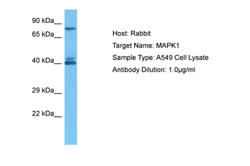
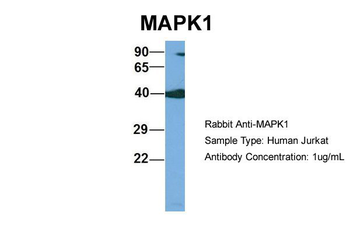
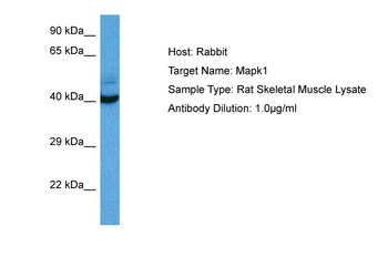
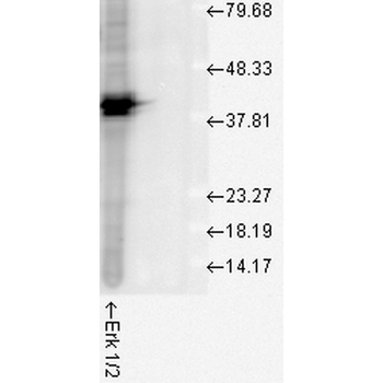
| Molecular Weight | 42kDa (ERK2). |
| Purification | Peptide Affinity Purified |
| Conjugation | Unconjugated |
Storage & Handling
−| Storage | Maintain refrigerated at 2-8°C for up to 2 weeks. For long term storage store at -20°C in small aliquots to prevent freeze-thaw cycles. |
|---|---|
| Buffer/Preservatives | PBS pH 7.4, 50% glycerol, 0.09% sodium azide. Storage buffer changes when conjugated. |
| Concentration | 1 mg/ml |
| Expiration Date | 12 months from date of receipt. |
| Disclaimer | For research use only |
Alternative Names
−ERK1, ERK2, ERT1, ERT2, MAP kinase 1, MAP kinase 2, MAPK1, MAPK2, MAPK3, p38, p40, p41, p41mapk, p42 MAPK, p44 ERK1, p44 MAPK, PRKM1, PRKM2, PRKM3
Similar Products
−- Item 1 of 16
Phospho-ERK1/2 (Thr202 + Tyr204) Rabbit Polyclonal Antibody [orb5178]
FC, ICC, IF, IHC-Fr, IHC-P
Bovine, Canine, Equine, Gallus, Guinea pig, Porcine, Rabbit
Human, Mouse, Rat
Rabbit
Polyclonal
Unconjugated
50 μl, 100 μl, 200 μl - Item 1 of 7
ERK1/2 Recombinant Rabbit Monoclonal Antibody [orb704524]
FC, ICC, IF, IHC-Fr, IHC-P, WB
Zebrafish
Human, Mouse, Rat
Rabbit
Recombinant
Unconjugated
50 μl, 100 μl, 25 μl - Item 1 of 5
ERK1/2 Mouse Monoclonal Antibody [orb500888]
FC, IF, IHC-Fr, IHC-P, WB
Mouse, Rat
Human, Mouse, Rat
Mouse
Monoclonal
Unconjugated
200 μg, 200 μl, 50 μl, 100 μl - Item 1 of 4
ERK1/2 Mouse Monoclonal Antibody [orb500875]
FC, IF, IHC-Fr, IHC-P, WB
Human
Human, Mouse, Rat
Mouse
Monoclonal
Unconjugated
200 μg, 200 μl, 50 μl, 100 μl - Item 1 of 6
MAPK1 Rabbit Polyclonal Antibody [orb584155]
IHC, WB
Bovine, Canine, Equine, Goat, Guinea pig, Mouse, Rabbit, Zebrafish
Human, Rat
Rabbit
Polyclonal
Unconjugated
100 μl


Quality Guarantee
Explore bioreagents carefree to elevate your research. All our products are rigorously tested for performance. If a product does not perform as described on its datasheet, our scientific support team will provide expert troubleshooting, a prompt replacement, or a refund. For full details, please see our Terms & Conditions and Buying Guide. Contact us at support@biorbyt.com.
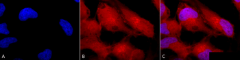
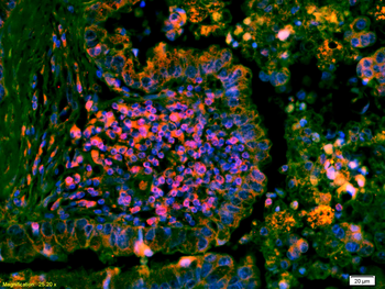
Immunocytochemistry/Immunofluorescence analysis using Rabbit Anti-Erk1/2 Polyclonal Antibody. Tissue: Cervical cancer cell line (HeLa). Species: Human. Fixation: 2% Formaldehyde for 20 min at RT. Primary Antibody: Rabbit Anti-Erk1/2 Polyclonal Antibody at 1:100 for 12 hours at 4°C. Secondary Antibody: APC Goat Anti-Rabbit (red) at 1:200 for 2 hours at RT. Counterstain: DAPI (blue) nuclear stain at 1:40000 for 2 hours at RT. Localization: Cytoplasm. Nucleus. Magnification: 100x. (A) DAPI (blue) nuclear stain. (B) Anti-Erk1/2 Antibody. (C) Composite.
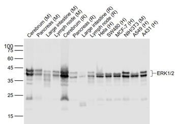
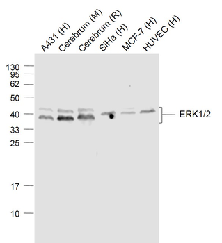
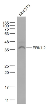
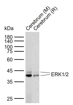
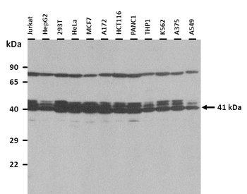
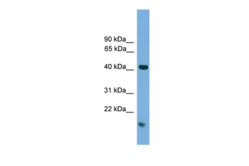
Western blot analysis of Human Cell line lysates showing detection of ERK1 protein using Rabbit Anti-ERK1 Polyclonal Antibody. Load: 15 μgprotein. Block: 1.5% BSA for 30 minutes at RT. Primary Antibody: Rabbit Anti-ERK1 Polyclonal Antibody at 1:1000 for 2 hours at RT. Secondary Antibody: Donkey Anti-Rabbit IgG: HRP for 1 hour at RT.
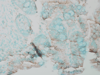
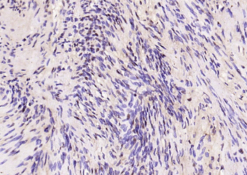
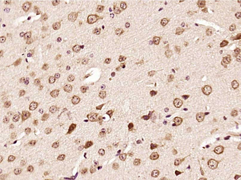
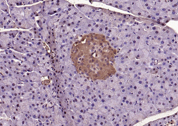
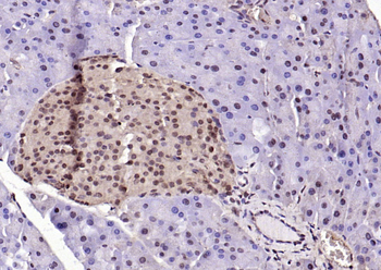
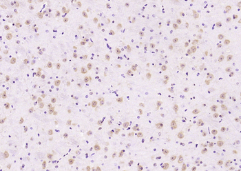
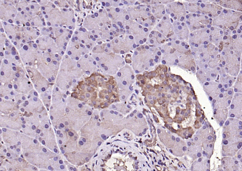
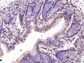
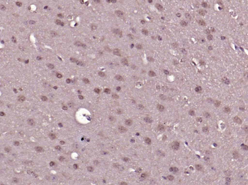
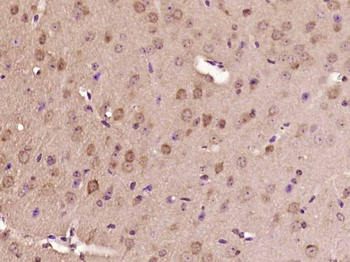
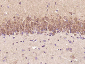
Immunohistochemistry analysis using Rabbit Anti-ERK1 Polyclonal Antibody. Tissue: Inflamed colon. Species: Mouse. Fixation: Formalin. Primary Antibody: Rabbit Anti-ERK1 Polyclonal Antibody at 1:25000 for 12 hours at 4°C. Secondary Antibody: Biotin Goat Anti-Rabbit at 1:2000 for 1 hour at RT. Counterstain: Methyl Green at 200uL for 2 min at RT.
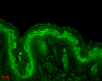
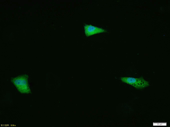
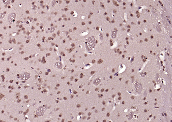
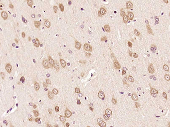
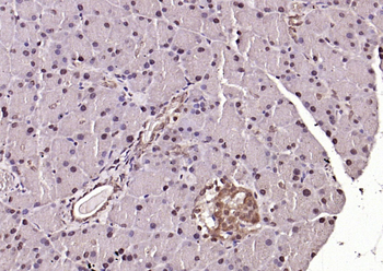
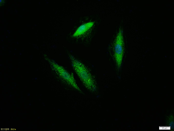
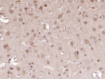
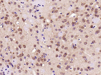
Immunohistochemistry analysis using Rabbit Anti-ERK1 Polyclonal Antibody. Tissue: backskin. Species: Mouse. Fixation: Bouin's Fixative Solution. Primary Antibody: Rabbit Anti-ERK1 Polyclonal Antibody at 1:100 for 1 hour at RT. Secondary Antibody: FITC Goat Anti-Rabbit (green) at 1:50 for 1 hour at RT. Localization: Cytoplasm.
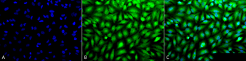
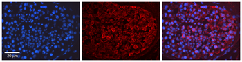
Immunocytochemistry/Immunofluorescence analysis using Rabbit Anti-Erk1/2 Polyclonal Antibody. Tissue: Cervical cancer cell line (HeLa). Species: Human. Fixation: 2% Formaldehyde for 20 min at RT. Primary Antibody: Rabbit Anti-Erk1/2 Polyclonal Antibody at 1:100 for 12 hours at 4°C. Secondary Antibody: FITC Goat Anti-Rabbit (green) at 1:200 for 2 hours at RT. Counterstain: DAPI (blue) nuclear stain at 1:40000 for 2 hours at RT. Localization: Cytoplasm. Nucleus. Magnification: 20x. (A) DAPI (blue) nuclear stain. (B) Anti-Erk1/2 Antibody. (C) Composite.
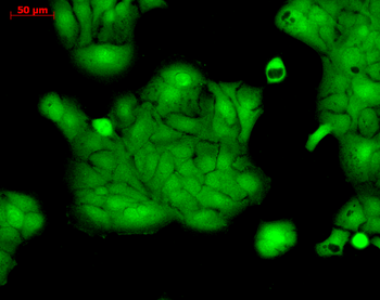
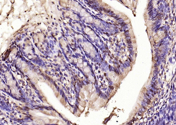
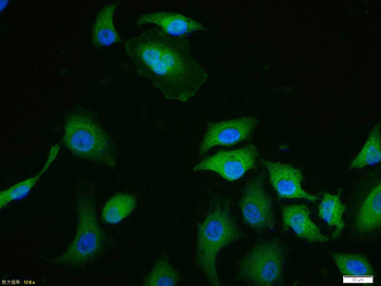
Immunocytochemistry/Immunofluorescence analysis using Rabbit Anti-ERK1 Polyclonal Antibody. Tissue: HaCaT cells. Species: Human. Fixation: Cold 100% methanol at -20°C for 10 minutes. Primary Antibody: Rabbit Anti-ERK1 Polyclonal Antibody at 1:100 for 12 hours at 4°C. Secondary Antibody: FITC Goat Anti-Rabbit at 1:50 for 1-2 hours at RT in dark. Localization: Cytoplasm. Nucleus.
Quick Database Links
UniProt Details
− No UniProt data available
NCBI Gene Details
− No NCBI Gene data available
NCBI Reference Sequences
−Associated Accession Numbers
Curated reference sequences for the gene transcript and protein product| Protein | NP_059043.1 |
|---|
Documents Download
Datasheet
Product Information
Request a Document
Protocol Information
WB
Western Blot (IB, immunoblot)
IHC
Immunohistochemistry
FC
Flow Cytometry
IF
Immunofluorescence
ICC
Immunocytochemistry
Erk1/2 Antibody (orb1822436)

- 0.0
Based on 0 reviews
Participating in our Biorbyt product reviews program enables you to support fellow scientists by sharing your firsthand experience with our products.
Login to Submit a ReviewAvailable Sizes
Select a size below
Free Secondary Antibody (20 ul)0/0
Please add an antibody product to your cart first.