You have no items in your shopping cart.
Description
Research Area
Signal Transduction
Images & Validation
−Item 1 of 3
| Tested Applications | IF, IHC-P, WB |
|---|---|
| Dilution Range | IF - 1:25, WB - 1:2000, IHC-P - 1:25 |
| Reactivity | Human |
Key Properties
−| Host | Mouse |
|---|---|
| Clonality | Monoclonal |
| Isotype | IgG1,k |
| Clone No. | B3662EV994X72X45 |
| Immunogen | This FGFR1 antibody is generated from a mouse immunized with a KLH conjugated synthetic peptide between 806-842 amino acids from the C-terminal region of human FGFR1. |
| Target | FGFR1 |
| Molecular Weight | 91868 Da |
| Conjugation | Unconjugated |
Storage & Handling
−| Storage | Maintain refrigerated at 2-8°C for up to 2 weeks. For long term storage store at -20°C in small aliquots to prevent freeze-thaw cycles |
|---|---|
| Form/Appearance | Purified monoclonal antibody supplied in PBS with 0.09% (W/V) sodium azide. This antibody is purified through a protein G column, followed by dialysis against PBS. |
| Expiration Date | 12 months from date of receipt. |
| Disclaimer | For research use only |
Alternative Names
−Fibroblast growth factor receptor 1, FGFR-1, Basic fibroblast growth factor receptor 1, BFGFR, bFGF-R-1, Fms-like tyrosine kinase 2, FLT-2, N-sam, Proto-oncogene c-Fgr, CD331, FGFR1, BFGFR, CEK, FGFBR, FLG, FLT2, HBGFR
Similar Products
−- Item 1 of 3
FGFR1 Mouse Monoclonal Antibody [orb1474017]
IF, IHC, WB
Human
Mouse
Monoclonal
Unconjugated
200 μl, 100 μl, 50 μl, 30 μl - Item 1 of 3
FOP Rabbit Polyclonal Antibody [orb215039]
IF, IHC, WB
Bovine, Canine, Human, Mouse, Porcine, Rat
Rabbit
Polyclonal
Unconjugated
30 μl, 100 μl, 200 μl, 50 μl - Item 1 of 2
Mouse Fgfr1 Antibody (C-term) [orb33072]
IHC-P, WB
Human, Mouse
Rabbit
Polyclonal
Unconjugated
50 μl, 100 μl - Item 1 of 2
FGFR1 Rabbit Polyclonal Antibody [orb216106]
IF, WB
Human
Rabbit
Polyclonal
Unconjugated
30 μl, 100 μl, 200 μl, 50 μl - Item 1 of 2
FGFR1 Rabbit Polyclonal Antibody [orb393289]
IHC, WB
Bovine, Human, Monkey, Mouse, Porcine, Rabbit, Rat
Rabbit
Polyclonal
Unconjugated
200 μl, 30 μl, 100 μl, 50 μl
Quality Guarantee
Explore bioreagents carefree to elevate your research. All our products are rigorously tested for performance. If a product does not perform as described on its datasheet, our scientific support team will provide expert troubleshooting, a prompt replacement, or a refund. For full details, please see our Terms & Conditions and Buying Guide. Contact us at support@biorbyt.com.
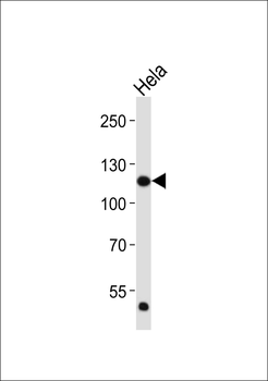
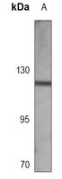
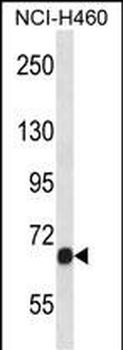
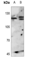
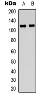
Western blot analysis of lysate from Hela cell line, using FGFR1 Antibody (C-term). Diluted at 1:2000. A goat anti-mouse IgG H&L (HRP) at 1: 3000 dilution was used as the secondary antibody. Lysate at 20 μg.
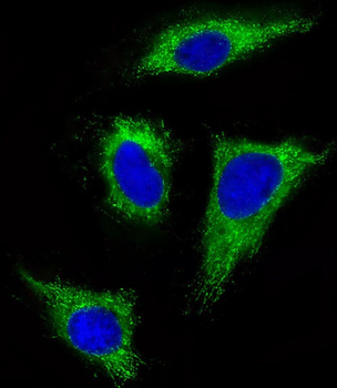
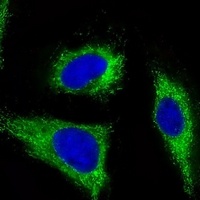
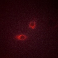
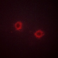
Immunofluorescent analysis of 4% paraformaldehyde-fixed, 0.1% Triton X-100 permeabilized HeLa (human cervical epithelial adenocarcinoma cell line) cells labeling FGFR1 at 1/25 dilution, followed by Dylight 488-conjugated goat anti-mouse IgG secondary antibody at 1/200 dilution (green). Immunofluorescence image showing cytoplasm staining on HeLa cell line. The nuclear counter stain is DAPI (blue).
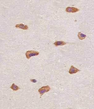
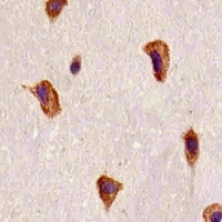
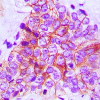
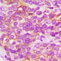
Staining FGFR1 in human brain tissue sections by Immunohistochemistry (IHC-P - paraformaldehyde-fixed, paraffin-embedded sections). Tissue was fixed with formaldehyde and blocked with 3% BSA for 0.5 hour at room temperature; antigen retrieval was by heat mediation with a citrate buffer (pH6). Samples were incubated with primary antibody (1/25) for 1 hours at 37°C. A undiluted biotinylated goat polyvalent antibody was used as the secondary antibody.
Quick Database Links
Gene Symbol
FGFR1
UniProt
UniProt Details
− No UniProt data available
Documents Download
Datasheet
Product Information
Request a Document
Protocol Information
WB
Western Blot (IB, immunoblot)
IHC-P
Immunohistochemistry Paraffin
IF
Immunofluorescence
FGFR1 Antibody (C-term) (orb1926703)

- 0.0
Based on 0 reviews
Participating in our Biorbyt product reviews program enables you to support fellow scientists by sharing your firsthand experience with our products.
Login to Submit a ReviewAvailable Sizes
Select a size below
Choose Conjugation or Carrier Free Version
Free Secondary Antibody (20 ul)0/0
Please add an antibody product to your cart first.