You have no items in your shopping cart.
Description
Research Area
Metabolism Research
Images & Validation
−Item 1 of 6
| Tested Applications | IF, IHC-P, WB |
|---|---|
| Dilution Range | IF - 1:10-50, WB - 1:1000, IHC-P - 1:10-50 |
| Reactivity | Human |
Key Properties
−| Host | Rabbit |
|---|---|
| Clonality | Polyclonal |
| Isotype | Rabbit IgG |
| Immunogen | This GAPDH antibody is generated from rabbits immunized with a KLH conjugated synthetic peptide between 62-91 amino acids from the N-terminal region of human GAPDH. Antigen Region: 62-91 aa. |
| Target | GAPDH {ECO:0000303|PubMed:2987855, ECO:0000312|HGNC:HGNC:4141} |
| Molecular Weight | 36053 Da |
| Conjugation | Unconjugated |
Storage & Handling
−| Storage | Maintain refrigerated at 2-8°C for up to 2 weeks. For long term storage store at -20°C in small aliquots to prevent freeze-thaw cycles |
|---|---|
| Form/Appearance | Purified polyclonal antibody supplied in PBS with 0.09% (W/V) sodium azide. This antibody is prepared by Saturated Ammonium Sulfate (SAS) precipitation followed by dialysis against PBS. |
| Expiration Date | 12 months from date of receipt. |
| Disclaimer | For research use only |
Alternative Names
−Glyceraldehyde-3-phosphate dehydrogenase, GAPDH, Peptidyl-cysteine S-nitrosylase GAPDH, 2699-, GAPDH, GAPD
Similar Products
−- Item 1 of 2
GAPDH Rabbit Polyclonal Antibody [orb213967]
IF, WB
Bovine, Canine, Gallus, Human, Monkey, Mouse, Porcine, Rabbit, Rat
Rabbit
Polyclonal
Unconjugated
30 μl, 100 μl, 200 μl, 50 μl - Item 1 of 1
GAPDS Rabbit Polyclonal Antibody [orb234857]
WB
Human, Monkey, Mouse, Rat
Rabbit
Polyclonal
Unconjugated
30 μl, 100 μl, 200 μl, 50 μl
Quality Guarantee
Explore bioreagents carefree to elevate your research. All our products are rigorously tested for performance. If a product does not perform as described on its datasheet, our scientific support team will provide expert troubleshooting, a prompt replacement, or a refund. For full details, please see our Terms & Conditions and Buying Guide. Contact us at support@biorbyt.com.
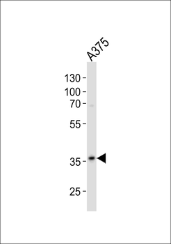
Western blot analysis of lysate from A375 cell line, using GAPDH Antibody (N-term). Diluted at 1:500. A goat anti-rabbit IgG H&L (HRP) at 1:10000 dilution was used as the secondary antibody. Lysate at 20 ug.
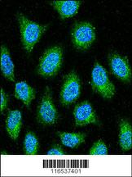
GAPDH Antibody (N-term) confocal immunofluorescent analysis with Hela cell. 0.025 mg/ml primary antibody was followed by FITC-conjugated goat anti-rabbit lgG (whole molecule). FITC emits green fluorescence. DAPI was used to stain the cell nuclear (blue).
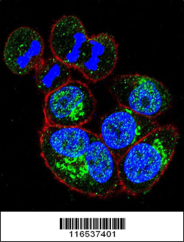
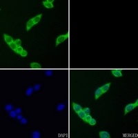
Confocal immunofluorescent analysis of GAPDH Antibody (N-term) with Hela cell followed by Alexa Fluor 488-conjugated goat anti-rabbit lgG (green). Actin filaments have been labeled with Alexa Fluor 555 phalloidin (red).DAPI was used to stain the cell nuclear (blue).
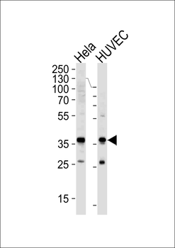
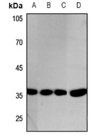
Western blot analysis of lysates from Hela, HUVEC cell line (from left to right), using GAPDH Antibody (N-term).Diluted at 1:1000 at each lane. A goat anti-rabbit IgG H&L (HRP) at 1:5000 dilution was used as the secondary antibody. Lysates at 35 ug per lane.
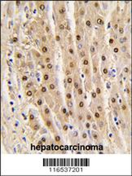
Formalin-fixed and paraffin-embedded human hepatocarcinoma tissue reacted with GAPDH antibody (N-term), which was peroxidase-conjugated to the secondary antibody, followed by DAB staining. This data demonstrates the use of this antibody for immunohistochemistry; clinical relevance has not been evaluated.
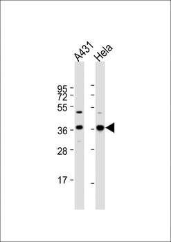
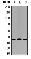
All lanes: Anti-GAPDH Antibody (N-term) at 1:1000 dilution. Lane 1: A431 whole cell lysate. Lane 2: Hela whole cell lysate. Lysates/proteins at 20 µg per lane. Secondary Goat Anti-Rabbit IgG, (H+L), Peroxidase conjugated at 1/10000 dilution. Predicted band size: 36 kDa. Blocking/Dilution buffer: 5% NFDM/TBST.
Quick Database Links
Gene Symbol
GAPDH {ECO:0000303|PubMed:2987855, ECO:0000312|HGNC:HGNC:4141}
UniProt
RefSeq (Protein):NP_001243728.1, NP_002037.2
UniProt Details
− No UniProt data available
NCBI Reference Sequences
−Associated Accession Numbers
Curated reference sequences for the gene transcript and protein product| Protein | NP_001243728.1, NP_002037.2 |
|---|
Documents Download
Datasheet
Product Information
Request a Document
Protocol Information
WB
Western Blot (IB, immunoblot)
IHC-P
Immunohistochemistry Paraffin
IF
Immunofluorescence
Filter by Applications
Filter by Species
Jie Wu 1, Hongya Wu 2, Bin Zhou 2, Xuefei He 1, Zhengjia Kang 1, Tiantian Chen 1, Gaoqin Liu 1, Peirong Lu 1 Effect of Topical Application of Dexamethasone on CC Chemokine Receptor 3 Expression in Human Pterygium J Ocul Pharmacol Ther, (2025)
Applications
WB
Reactivity
Human
GAPDH Antibody (N-term) (orb1928806)

- 0.0
Based on 0 reviews
Participating in our Biorbyt product reviews program enables you to support fellow scientists by sharing your firsthand experience with our products.
Login to Submit a ReviewAvailable Sizes
Select a size below
Choose Conjugation or Carrier Free Version
Free Secondary Antibody (20 ul)0/0
Please add an antibody product to your cart first.