You have no items in your shopping cart.
Description
Research Area
Neuroscience
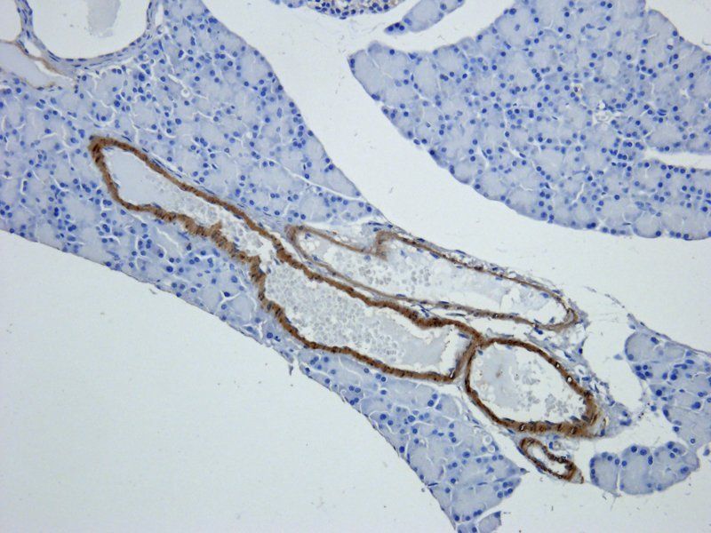
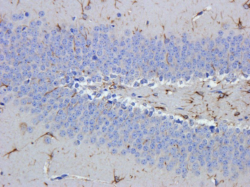
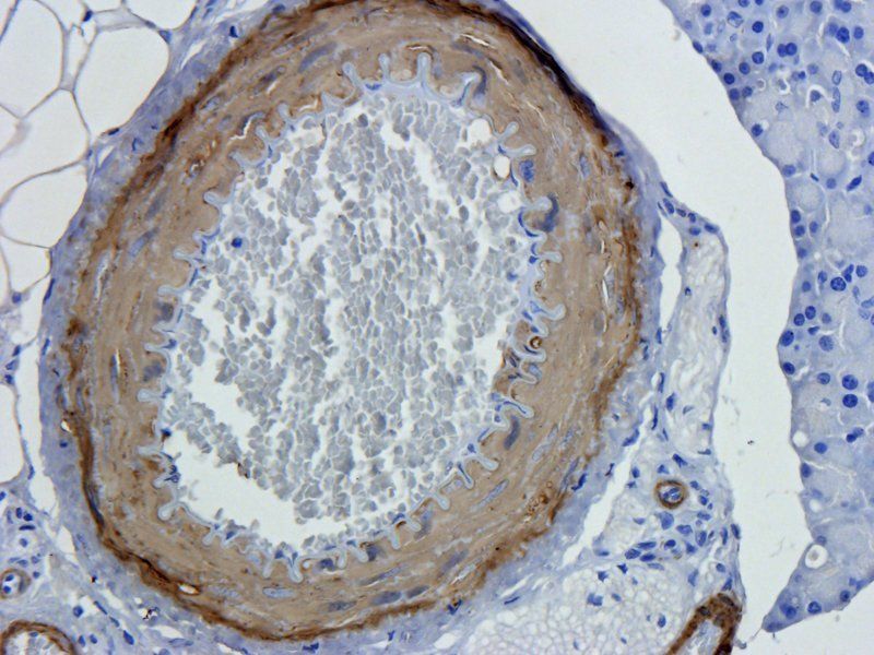
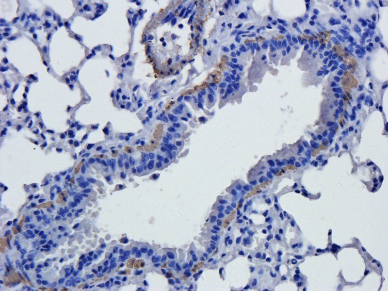
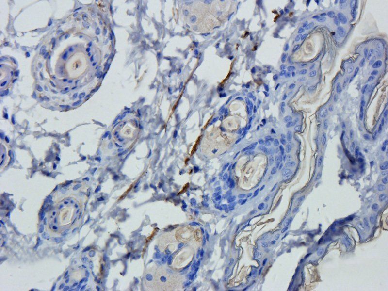
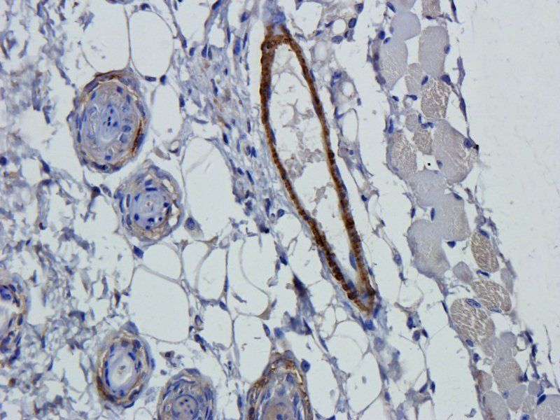
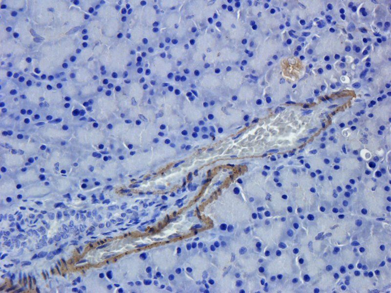
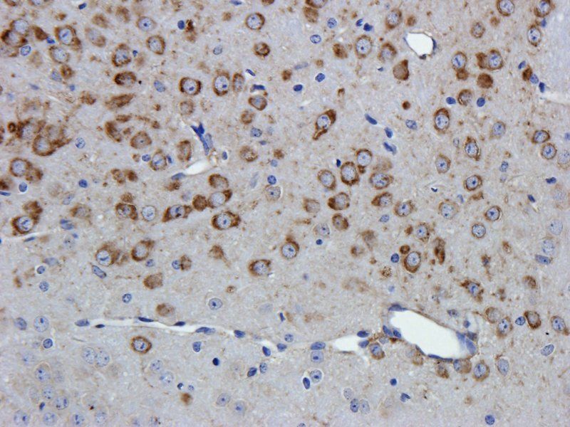
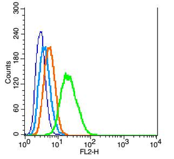
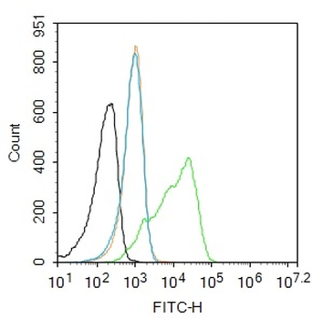
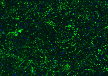
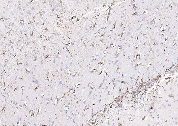
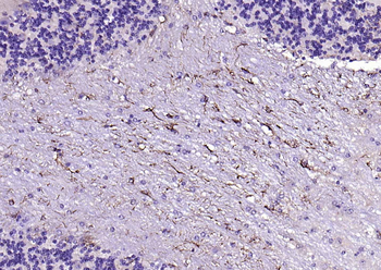
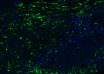
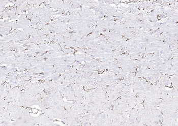
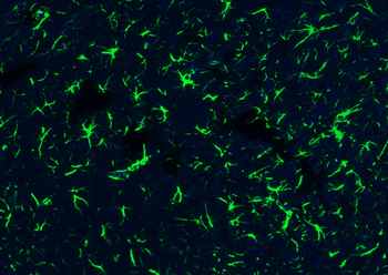
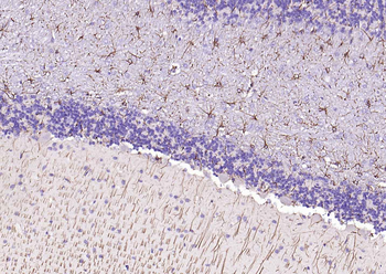
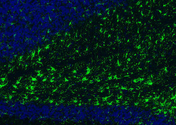
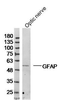
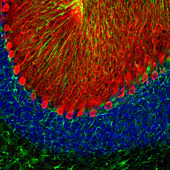
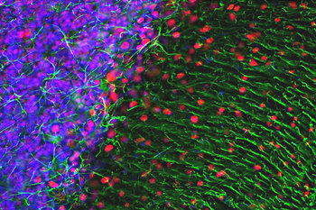
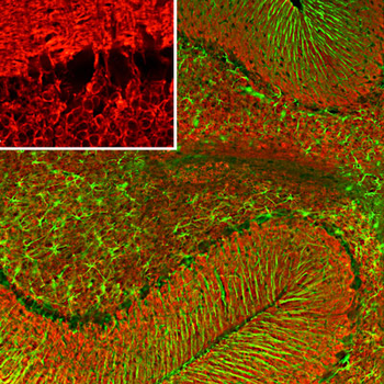
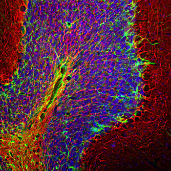
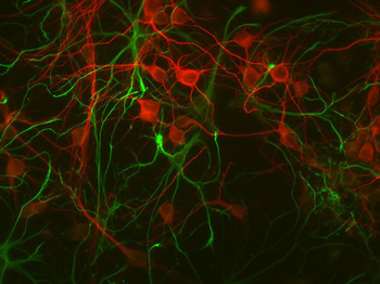
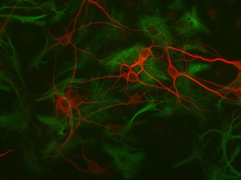
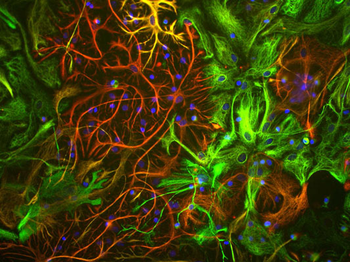
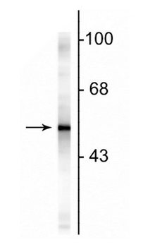
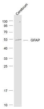
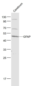
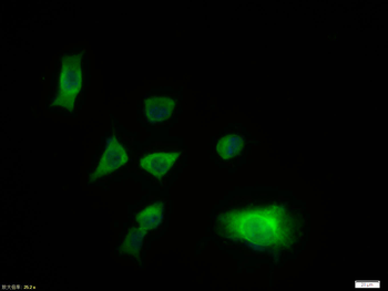
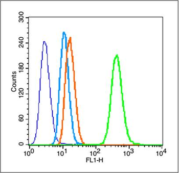
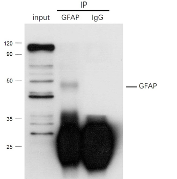
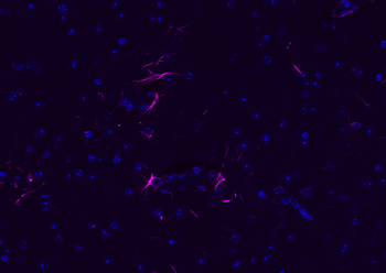
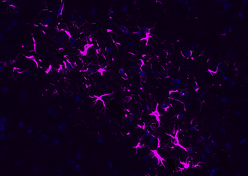
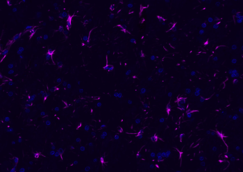
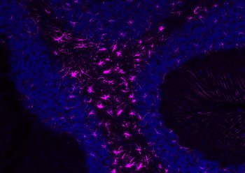
Images & Validation
−
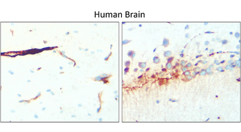
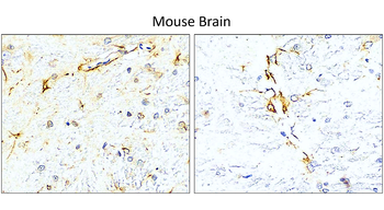

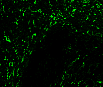
Item 1 of 8
| Tested Applications | IF, IHC-Fr, IHC-P, WB |
|---|---|
| Dilution Range | Western Blot 1:1000-1:2000, Immunofluorescence 1:200 – 1:500, Immunohistochemistry (Frozen) 1:100 – 1:300, Immunohistochemistry (Paraffin) 1:100 – 1:300 |
| Reactivity | Human, Mouse, Rat |
Key Properties
−| Antibody Type | Primary Antibody |
|---|---|
| Host | Rabbit |
| Clonality | Polyclonal |
| Isotype | Rabbit-IgG |
| Immunogen | Synthetic peptide / encompassing a sequence within the C-terminus region. |
| Target | GFAP |
| Conjugation | Unconjugated |
Storage & Handling
−| Storage | Maintain refrigerated at 2-8°C for up to 2 weeks. For long term storage store at -20°C in small aliquots to prevent freeze-thaw cycles. |
|---|---|
| Buffer/Preservatives | 100mM Tris Glycine, 1% rAlbumin, 20% Glycerol (pH7). 0.025% ProClin 300 was added as a preservative |
| Expiration Date | 12 months from date of receipt. |
| Disclaimer | For research use only |
Similar Products
−- Item 1 of 15
GFAP Rabbit Polyclonal Antibody [orb10706]
ELISA, ICC, IF, IHC-P, WB
Human, Mouse, Rat
Rabbit
Polyclonal
Unconjugated
100 μg - Item 1 of 12
GFAP Rabbit Polyclonal Antibody [orb500829]
FC, IF, IHC-Fr, IHC-P
Bovine, Canine, Porcine, Rabbit, Sheep
Human, Mouse, Rat
Rabbit
Polyclonal
Unconjugated
50 μl, 100 μl, 200 μl - Item 1 of 8
Glial Fibrillary Acidic Proetin (GFAP) Antibody [orb319549]
ELISA, ICC, IHC, WB
Bovine, Equine, Human, Mouse, Rat
Rabbit
Polyclonal
Unconjugated
100 μl - Item 1 of 5
GFAP Rabbit Polyclonal Antibody [orb221420]
FC, WB
Bovine, Gallus, Porcine, Rabbit, Sheep
Human, Mouse, Rat
Rabbit
Polyclonal
Unconjugated
- Item 1 of 4
GFAP Rabbit Polyclonal Antibody (Cy5) [orb913879]
FC, IF
Bovine, Canine, Porcine, Rabbit, Sheep
Human, Mouse, Rat
Rabbit
Polyclonal
Cy5
100 μl


Quality Guarantee
Explore bioreagents carefree to elevate your research. All our products are rigorously tested for performance. If a product does not perform as described on its datasheet, our scientific support team will provide expert troubleshooting, a prompt replacement, or a refund. For full details, please see our Terms & Conditions and Buying Guide. Contact us at support@biorbyt.com.
Quick Database Links
Gene Symbol
GFAP
Protocol Information
WB
Western Blot (IB, immunoblot)
IHC-P
Immunohistochemistry Paraffin
IHC-Fr
Immunohistochemistry Frozen
IF
Immunofluorescence
Available Sizes
Select a size below
Choose Conjugation or Carrier Free Version
Free Secondary Antibody (20 ul)0/0
Please add an antibody product to your cart first.