You have no items in your shopping cart.
Description
Images & Validation
−Item 1 of 5
| Tested Applications | ELISA, IF, IHC, Multiplex Assay, WB |
|---|---|
| Dilution Range | ELISA: 1:6,000 - 1:30,000, IHC: 0.5 mg/ml - 5 µg/ml, WB: 1:500 - 1:2,000 |
| Reactivity | Human |
| Application Notes |
Key Properties
−| Antibody Type | Primary Antibody |
|---|---|
| Host | Rabbit |
| Clonality | Polyclonal |
| Isotype | IgG |
| Immunogen | This affinity purified antibody was produced from monospecific rabbit serum by repeated immunizations with a synthetic peptide corresponding to an internal region near amino acids 30-60 of human Gli-3 protein. |
| Target | GLI3 |
| Purity | This affinity-purified antibody is directed against human Gli-3 protein. The product was affinity purified from monospecific antiserum by immunoaffinity purification. A BLAST analysis was used to suggest cross reactivity with Gli-3 from human, chimpanzee, squirrel monkey, Xenopus laevis, chicken, dog and quail based on 100% sequence homology with the immunogen. Reactivity is also expected against homologues from mouse (94%) and rat (88%) based on partial homology. Reactivity with Gli-3 from other sources is not known. |
| Conjugation | Unconjugated |
Storage & Handling
−| Storage | Store vial at -20° C prior to opening. Aliquot contents and freeze at -20° C or below for extended storage. Avoid cycles of freezing and thawing. Centrifuge product if not completely clear after standing at room temperature. This product is stable for several weeks at 4° C as an undiluted liquid. Dilute only prior to immediate use. |
|---|---|
| Form/Appearance | Liquid (sterile filtered) |
| Buffer/Preservatives | Preservative: 0.01% (w/v) Sodium Azide. Stabilizer: None; Buffer: 0.02 M Potassium Phosphate, 0.15 M Sodium Chloride, pH 7.2 |
| Concentration | 1.0 mg/mL |
| Expiration Date | 12 months from date of receipt. |
| Dry Ice Shipping | Please note: This product requires shipment on dry ice. A dry ice surcharge will apply. |
| Disclaimer | For research use only |
Alternative Names
−Rabbit anti-GLI-3 antibody, Transcriptional activator GLI3, Gli 3, GLI3 form of 190 kDa, GLI3 form of 83 kDa
Similar Products
−- Item 1 of 2
GLI-3 rabbit pAb Antibody [orb768436]
ELISA, IF, IHC
Human, Mouse, Rat
Polyclonal
Unconjugated
50 μl, 100 μl - Item 1 of 2
Gli3 Rabbit Polyclonal Antibody [orb157158]
IF, IHC-Fr, IHC-P
Bovine, Equine, Gallus, Human, Rabbit, Sheep
Mouse, Rat
Rabbit
Polyclonal
Unconjugated
50 μl, 100 μl, 200 μl - Item 1 of 1
GLI3 Rabbit Polyclonal Antibody [orb2952790]
ELISA, IHC, WB
Human, Mouse, Rat
Rabbit
Polyclonal
Unconjugated
100 μg, 50 μg - Item 1 of 2
GLI3 Rabbit Polyclonal Antibody [orb574133]
IHC
Bovine, Canine, Equine, Guinea pig, Human, Mouse, Rabbit, Rat, Zebrafish
Rabbit
Polyclonal
Unconjugated
100 μl - Item 1 of 1
GLI3 Specific Rabbit Polyclonal Antibody [orb395084]
ELISA, WB
Human, Mouse, Rat
Rabbit
Polyclonal
Unconjugated
50 μg, 100 μg

Quality Guarantee
Explore bioreagents carefree to elevate your research. All our products are rigorously tested for performance. If a product does not perform as described on its datasheet, our scientific support team will provide expert troubleshooting, a prompt replacement, or a refund. For full details, please see our Terms & Conditions and Buying Guide. Contact us at support@biorbyt.com.
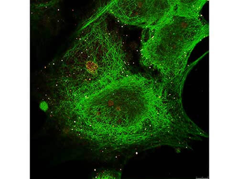
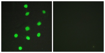
Immunofluorescence Microscopy of Rabbit anti-Gli-3 antibody. Tissue: MCF-7 cell. Antigen retrieval: not required. Primary antibody: Gli-3 antibody and Anti-alpha-Tubulin at 5 µg/ml for 1 h at RT. Secondary antibody: Fluorescein secondary antibody at 1:10000 for 45 min at RT. Localization: Gli-3 is nuclear. Staining: Gli-3 staining as red fluorescent signal and Anti-alpha-Tubulin staining as green fluorescent signal using STED.
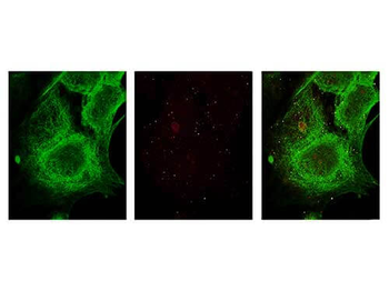
Immunofluorescence Microscopy of Rabbit anti-Gli-3 antibody. Tissue: MCF-7 cell. Antigen retrieval: not required. Primary antibody: Gli-3 antibody and Anti-alpha-Tubulin at 5 µg/ml for 1 h at RT. Secondary antibody: Fluorescein secondary antibody at 1:10000 for 45 min at RT. Localization: Gli-3 is nuclear. Staining: Image (1) shows alpha-Tubulin staining as green fluorescent signal. Image (2) shows Gli-3 staining as red fluorescent signal and Images (3) shows both antibodies fluorescing using STED microscopy.
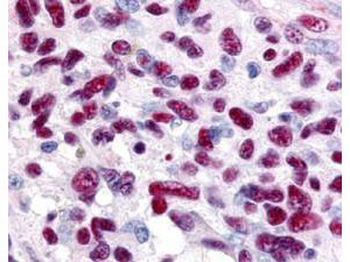
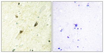
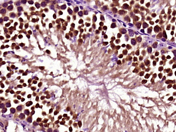
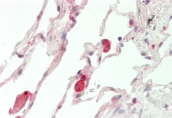
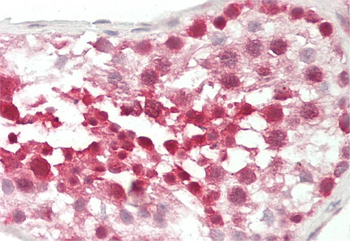
Immunohistochemistry of Rabbit anti-Gli-3 antibody. This image tissue: human glioblastoma. Specific staining was also noted in tissue from adrenal, brain, glioblastoma, colon, heart, kidney, lung, liver, skeletal muscle, ovary, pancreas, placenta, skin, spleen, stomach, testes, thymus, thyroid, tonsil and uterus. Fixation: formalin fixed paraffin embedded. Antigen retrieval: not required. Primary antibody: Gli-3 antibody at 0.625 µg/ml for 1 h at RT. Secondary antibody: Peroxidase rabbit secondary antibody at 1:10000 for 45 min at RT. Localization: Gli-3 is nuclear and smooth muscle. Staining: Gli-3 as precipitated red signal with hematoxylin purple nuclear counterstain.
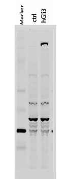
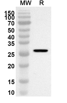
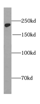
Western Blot of Rabbit anti-Gli-3 antibody. Lane 1: 50 kDa molecular weight marker. Lane 2: 293T cells transfected with CrkL-Flag. Lane 3: 293T cells transfected with human Gli-3. Load: 35 µg per lane. Primary antibody: Gli-3 antibody at 1:400 for overnight at 4°C. Secondary antibody: IRDye800™ rabbit secondary antibody at 1:10000 for 45 min at RT. Block: 5% BLOTTO overnight at 4°C. Predicted/Observed size: 170-190 kDa for hGli-3. Other band(s): Non specific background ~60kDa.
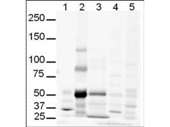
Western Blot of Rabbit anti-Gli-3 antibody. Lane 1: human brain whole cell lysate. Lane 2: human lung whole cell lysate. Lane 3: human spleen whole cell lysate. Lane 4: mouse brain whole cell lysate (p/n orb348715). Lane 5: mouse lung whole cell lysate (p/n orb692725). Load: 20 µg per lane. Primary antibody: Gli-3 antibody at 1:500 for overnight at 4°C. Secondary antibody: IRDye800™ rabbit secondary antibody at 1:10000 for 45 min at RT. Block: 5% BLOTTO overnight at 4°C. Predicted/Observed size: Isoforms at ~170-190kDa and ~80kDa. Lane 2 shows what may be truncated Gli-3 (~80kDa). Other band(s): The strong band at ~50 kDa is unknown.
Documents Download
Datasheet
Product Information
Request a Document
Protocol Information
WB
Western Blot (IB, immunoblot)
IHC
Immunohistochemistry
IF
Immunofluorescence
ELISA
Enzyme-linked Immunosorbent Assay (EIA)
GLI3 Antibody (orb345478)

- 0.0
Based on 0 reviews
Participating in our Biorbyt product reviews program enables you to support fellow scientists by sharing your firsthand experience with our products.
Login to Submit a ReviewAvailable Sizes
Select a size below
Choose Conjugation or Carrier Free Version
Free Secondary Antibody (20 ul)0/0
Please add an antibody product to your cart first.