You have no items in your shopping cart.
Featured
Description
Research Area
Epigenetics & Chromatin
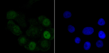
Images & Validation
−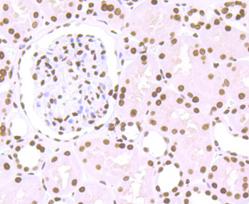
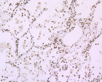
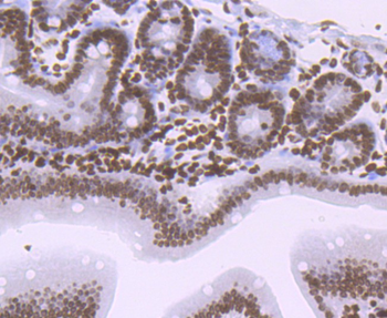
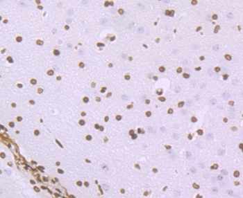

Item 1 of 8
| Tested Applications | FC, ICC, WB |
|---|---|
| Dilution Range | WB=1:500-2000, ICC/IF=1:50-200, Flow-Cyt=1:50-100 |
| Reactivity | Human, Mouse, Rat |
| Predicted Reactivity | Mouse, Rat |
Key Properties
−| Antibody Type | Primary Antibody |
|---|---|
| Host | Rabbit |
| Clonality | Recombinant |
| Isotype | IgG |
| Clone No. | B1E1 |
| Immunogen | KLH conjugated synthetic peptide derived from human Histone H1.2 |
| Target | H1-2 |
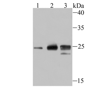
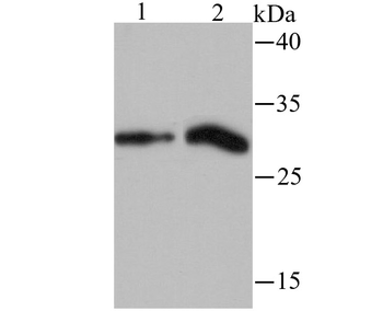
| Molecular Weight | 32 kDa |
| Purification | Affinity purified by Protein A |
| Conjugation | Unconjugated |
Storage & Handling
−| Storage | Maintain refrigerated at 2-8°C for up to 2 weeks. For long term storage store at -20°C in small aliquots to prevent freeze-thaw cycles. |
|---|---|
| Form/Appearance | Liquid |
| Buffer/Preservatives | 0.01M TBS (pH7.4) with 1% rAlbumin, 0.02% Proclin300 and 50% Glycerol. |
| Concentration | 1mg/ml |
| Expiration Date | 12 months from date of receipt. |
| Disclaimer | For research use only |
Alternative Names
−H1.2; H1C; H1F2; H1s-1; HIST1H1C; H12_HUMAN; H1-2; Histone H1c; Histone H1d; Histone H1s-1; H1.2 linker histone, cluster member; H1 histone family, member 2; histone 1, H1c; histone cluster 1, H1c; histone cluster 1 H1 family member c
Similar Products
−
HIST1H1C Rabbit Monoclonal Antibody [orb2989051]
WB
Human, Mouse, Rat
Rabbit
Monoclonal
Unconjugated
200 μl, 100 μl, 50 μl, 30 μl
Quality Guarantee
Explore bioreagents carefree to elevate your research. All our products are rigorously tested for performance. If a product does not perform as described on its datasheet, our scientific support team will provide expert troubleshooting, a prompt replacement, or a refund. For full details, please see our Terms & Conditions and Buying Guide. Contact us at support@biorbyt.com.
Quick Database Links
Gene Symbol
H1-2
UniProt
UniProt Details
− No UniProt data available
Protocol Information
WB
Western Blot (IB, immunoblot)
FC
Flow Cytometry
ICC
Immunocytochemistry
Available Sizes
Select a size below
Free Secondary Antibody (20 ul)0/0
Please add an antibody product to your cart first.