
You have no items in your shopping cart.

Protocol Optimization & Best Practices
Tips for Optimizing Your IF Staining
A comprehensive technical guide to immunofluorescence principles, troubleshooting non-specific signal, and multiplexing strategies.
Immunofluorescence (IF) is a powerful, fluorophore-based technique used to detect, localize, and track the subcellular distribution and dynamic movement of proteins and other biomolecules within cultured cells and tissue sections.
Unlike traditional chromogenic labeling methods used in Immunohistochemistry (IHC) or Immunocytochemistry (ICC) (such as DAB precipitation), IF utilizes distinct fluorophores that emit light at specific wavelengths when excited. By using multiple secondary antibodies or direct fluorophore conjugates with non-overlapping spectra, researchers can evaluate the co-localization of multiple target proteins simultaneously—generating high-resolution multiplexed data from a single sample.
Step-by-Step Protocol
Looking for detailed incubation times, fixative recipes, and wash protocols? Check out Biorbyt's official Immunofluorescence Protocol Guide for bench-ready instructions.
1. When to Use Immunofluorescence
IF is the preferred experimental approach under several specific biological conditions:
- Subcellular Localization: When mapping expression to specific organelle compartments (e.g., mitochondria, endoplasmic reticulum, nucleus, plasma membrane).
- Protein Co-Localization: When investigating physical interactions or spatial overlap between two or more target proteins within the same cell or tissue section.
- Dynamic Translocation: When tracking protein movement between cellular compartments (e.g., nuclear translocation upon receptor stimulation).
- Multiplexing Alternative: As a high-resolution fluorescent alternative to single-target chromogenic substrates like DAB or AEC.
2. Understanding Differences: IF vs. ICC vs. IHC
Understanding the distinction between these three terms relies on recognizing the sample type versus the detection mechanism:
- Immunohistochemistry (IHC): Refers specifically to staining intact tissue architecture—either formalin-fixed paraffin-embedded (FFPE) or frozen tissue sections. Detection can be chromogenic or fluorescent.
- Immunocytochemistry (ICC): Refers specifically to staining isolated cells—such as adherent cell cultures, suspension cells, or tissue imprints. Detection can be chromogenic or fluorescent.
- Immunofluorescence (IF): Refers to the fluorescent detection chemistry itself, which can be applied to either tissue sections (IHC-IF) or cultured cells (ICC-IF).
3. Direct vs. Indirect Immunofluorescence
Immunofluorescence can be performed using either direct or indirect labeling strategies:
- Direct IF: Uses a primary antibody that is directly conjugated to a fluorophore (e.g., FITC, Alexa Fluor 488, PE). This eliminates secondary antibody incubation steps, reducing overall protocol time and minimizing species cross-reactivity in multiplex panels. However, signal amplification is limited.
- Indirect IF: Uses an unconjugated primary antibody to bind the target antigen, followed by a fluorophore-conjugated secondary antibody raised against the host species of the primary. Multiple secondary antibodies bind each primary molecule, providing significant signal amplification for low-abundance targets.
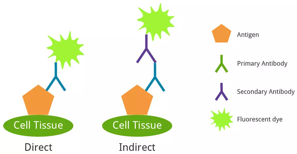
Direct IF (fluorophore-conjugated primary antibody) vs. Indirect IF (unconjugated primary + fluorophore-conjugated secondary antibody).
4. Troubleshooting Common IF Staining Issues
Identify and resolve common experimental errors that lead to non-specific staining, weak signal, or elevated background:
| Issue | Potential Causes & Solutions |
|---|---|
| Non-Specific Staining |
1. Fluorophore Spectral Overlap: Adjust excitation/emission optical filters or switch to fluorophores with non-overlapping emission spectra. 2. Primary Antibody Raised in Same Species as Sample: Use normal host serum (4%) matching the secondary antibody host species as a blocking agent. Alternatively, block primary antibodies with monovalent Fab fragments. 3. Antibody Aggregates: Microcentrifuge secondary antibodies prior to dilution to pellet protein aggregates. 4. Unreacted Fixative Aldehydes: Quench residual free aldehydes following formaldehyde/PFA fixation by washing with 0.1 M glycine or sodium borohydride (NaBH4). |
| Weak or Missing Signal |
1. Exposure & Intensity Settings: Increase excitation light intensity or prolong camera exposure times during acquisition. 2. Inadequate Permeabilization: Ensure internal organelles/nuclear targets are permeable by using 0.1%–0.3% Triton X-100 or Saponin in wash buffers. 3. Low Primary Antibody Concentration: Titrate primary antibody concentrations and extend primary incubation to overnight at 4°C. 4. Photobleaching: Avoid prolonged light exposure; utilize mounting media containing anti-fade agents (e.g., DABCO, Mowiol). |
| Elevated Background Signal |
1. Excessive Antibody Concentration: Reduce primary or secondary antibody concentration (titrate via serial dilutions). 2. Insufficient Blocking: Increase blocking incubation duration or switch to 4%–10% normal serum from the secondary antibody host species. 3. Endogenous Tissue Autofluorescence: Check unstained controls. Quench lipofuscin/heme autofluorescence using Sudan Black B staining or shift detection to far-red/near-IR channels (e.g., Alexa Fluor 647, Cy5). 4. Inadequate Washing: Increase wash duration and cycle frequency (3–5 washes of 5 minutes each using PBS with 0.05% Tween-20). |
5. Seven Best Practices for Optimizing IF
Include Essential Controls
Always include no-primary controls (secondary antibody only) to evaluate background binding, unstained controls to assess endogenous autofluorescence, and known positive/negative cell lines to verify target binding specificity.
Protect Reagents and Samples from Light
Fluorescently conjugated antibodies and stained coverslips are highly susceptible to photobleaching. Store conjugate vials in amber tubes or foil-wrapped containers, and perform all incubation and wash steps in dark or covered humidity chambers.
Select the Optimal Fixative
Cross-linking fixatives (e.g., 4% Paraformaldehyde/PFA) preserve cytoskeletal and nuclear structures well but require a secondary permeabilization step (Triton X-100). Precipitating organic solvents (e.g., cold Methanol or Acetone) simultaneously fix and permeabilize cells, making them ideal for frozen tissue sections and certain monoclonal antibodies.
Ensure Host Species Compatibility
Verify that your secondary antibody specifically recognizes the host species of your primary antibody (e.g., use a Goat anti-Mouse IgG secondary to detect a Mouse IgG primary). Avoid secondary antibodies raised in the same host species as your test tissue to prevent non-specific anti-IgG binding.
Optimize Multiplex Panel Design
When performing multi-color co-localization studies, select primary antibodies raised in distinct host species (e.g., Mouse primary + Rabbit primary). Use secondary antibodies that have been cross-adsorbed (pre-adsorbed) against other species to eliminate cross-species reactivity. Raising all secondary antibodies in the same host species (e.g., Goat anti-Mouse + Goat anti-Rabbit) ensures uniform blocking conditions.
Decide Between Direct vs. Indirect Staining
Weigh the advantages of each format: Direct IF offers faster protocols and fewer species conflicts for multiplexing, whereas Indirect IF provides higher sensitivity through secondary antibody signal amplification.
Eliminate Spectral Bleed-Through
Choose fluorophores with widely separated excitation and emission peaks (e.g., DAPI/Alexa Fluor 488/Alexa Fluor 568/Alexa Fluor 647). Configure microscope narrow-band pass filter cubes and acquire signals sequentially rather than simultaneously to prevent fluorophore bleed-through between channels.
Need Guidance on Fluorophore Selection?
Biorbyt’s scientific team can assist you with selecting pre-adsorbed secondary antibodies, fluorophore matching, and multiplex panel design.
Contact Scientific Support