You have no items in your shopping cart.
MET/HGFR Antibody
SKU: orb1440712
Description
Research Area
Cancer Biology, Signal Transduction
Images & Validation
−Item 1 of 3
| Tested Applications | IF, IHC-P, WB |
|---|---|
| Dilution Range | IF: 1:200, WB: 1:500~1000, IHC-P: 1:10~50 |
| Reactivity | Human, Mouse |
Key Properties
−| Antibody Type | Primary Antibody |
|---|---|
| Host | Mouse |
| Clonality | Monoclonal |
| Isotype | Mouse IgG1 |
| Clone No. | 6AT1785 |
| Target | MET |
| Molecular Weight | 155541 |
| Conjugation | Unconjugated |
Storage & Handling
−| Storage | Maintain refrigerated at 2-8°C for up to 2 weeks. For long term storage store at -20°C in small aliquots to prevent freeze-thaw cycles |
|---|---|
| Form/Appearance | Purified monoclonal antibody supplied in PBS with 0.09% (W/V) sodium azide. This antibody is purified through a protein G column, followed by dialysis against PBS. |
| Expiration Date | 12 months from date of receipt. |
| Disclaimer | For research use only |
Alternative Names
−Hepatocyte growth factor receptor, HGF receptor, HGF/SF receptor, Proto-oncogene c-Met, Scatter factor receptor, SF receptor, Tyrosine-protein kinase Met, MET
Similar Products
−- Item 1 of 4
MET/HGFR Antibody [orb1939458]
IF, IHC-P, WB
Human, Mouse, Rat
Mouse
Monoclonal
Unconjugated
100 μl, 50 μl - Item 1 of 3
- Item 1 of 1
Bi-Phospho-MET/HGFR(Y1234/Y1235) Antibody [orb1927589]
WB
Human
Mouse
Monoclonal
Unconjugated
100 μl, 50 μl - Item 1 of 3
- Item 1 of 4
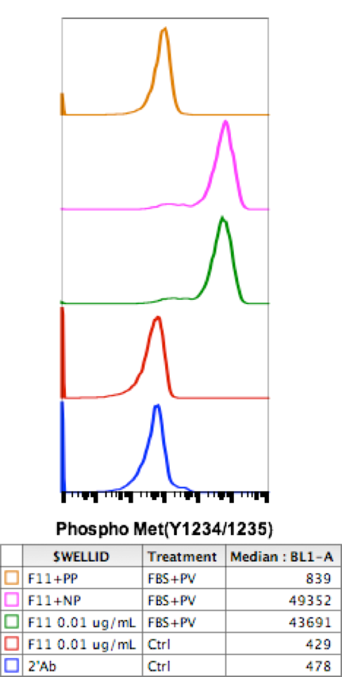
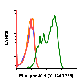
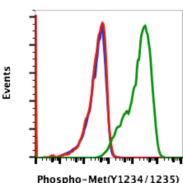
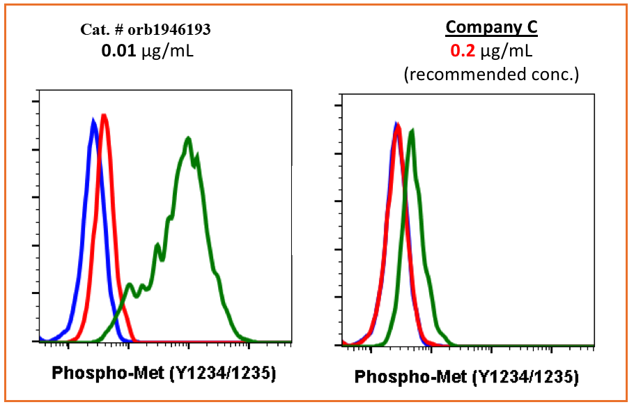
Phospho-MET (Tyr1234/1235) (6F11) rabbit mAb Antibody [orb1946193]
FC
Human, Mouse
Rabbit
Monoclonal
Unconjugated
200 μl, 20 μl


Quality Guarantee
Explore bioreagents carefree to elevate your research. All our products are rigorously tested for performance. If a product does not perform as described on its datasheet, our scientific support team will provide expert troubleshooting, a prompt replacement, or a refund. For full details, please see our Terms & Conditions and Buying Guide. Contact us at support@biorbyt.com.
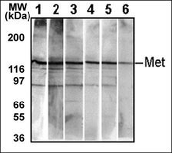
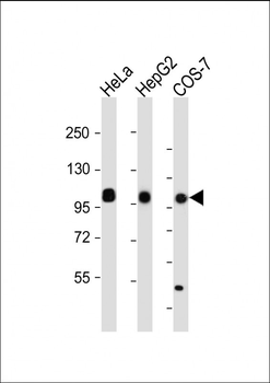
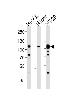
Detection of endogenous Met in HepG2 cell line. 10 μg/lane of HepG2 cell lysate was used to examine the expression of human Met. Lanes 1-5 represent Abgent's different anti-Met monoclonal antibodies. Lane 6 represents auto-phosohorylated-Met in HepG2 cell line detected by anti-phospho-Met Mab.
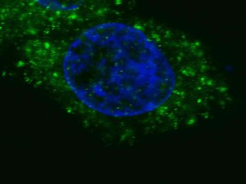
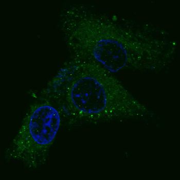
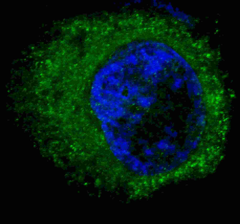
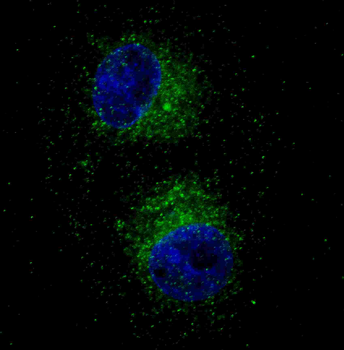
Fluorescent confocal image of HepG2 cells stained with MET/HGFR Antibody. HepG2 cells were fixed with 4% PFA (20 min), permeabilized with Triton X-100 (0.2%, 30 min). Cells were then incubated with MET/HGFR primary antibody (1:200, 2 h at room temperature). For secondary antibody, Alexa Fluor 488 conjugated donkey anti-mouse antibody (green) was used (1:1000, 1h). Nuclei were counterstained with Hoechst 33342 (blue) (10 μg/ml, 5 min).
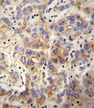
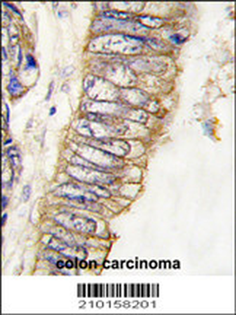
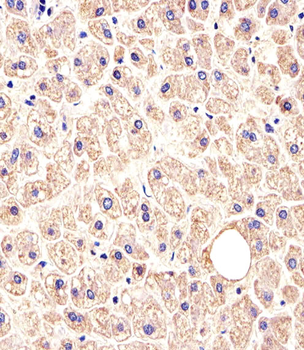
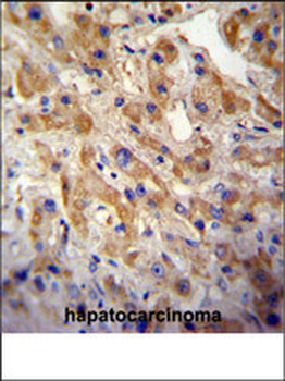
MET/HGFR Antibody immunohistochemistry analysis in formalin fixed and paraffin embedded human hepatocarcinoma followed by peroxidase conjugation of the secondary antibody and DAB staining. This data demonstrates the use of the MET/HGFR Antibody for immunohistochemistry. Clinical relevance has not been evaluated.
Quick Database Links
UniProt Details
− No UniProt data available
NCBI Reference Sequences
−Associated Accession Numbers
Curated reference sequences for the gene transcript and protein product| Protein | NP_000236.2, NP_001120972.1 |
|---|
Documents Download
Datasheet
Product Information
Request a Document
Protocol Information
WB
Western Blot (IB, immunoblot)
IHC-P
Immunohistochemistry Paraffin
IF
Immunofluorescence
MET/HGFR Antibody (orb1440712)

- 0.0
Based on 0 reviews
Participating in our Biorbyt product reviews program enables you to support fellow scientists by sharing your firsthand experience with our products.
Login to Submit a ReviewAvailable Sizes
Select a size below