You have no items in your shopping cart.
NEK2 Antibody (Center)
SKU: orb1788313
Description
Images & Validation
−Item 1 of 5
| Tested Applications | IF, IHC-P, WB |
|---|---|
| Dilution Range | IF: 1:25, WB: 1:1000, IHC-P: 1:100, IHC-P: 1:100, IHC-P: 1:100 |
| Reactivity | Human, Mouse |
Key Properties
−| Antibody Type | Primary Antibody |
|---|---|
| Host | Rabbit |
| Clonality | Polyclonal |
| Isotype | Rabbit IgG |
| Immunogen | 396-426 aa |
| Target | This NEK2 antibody is generated from rabbits immunized with a KLH conjugated synthetic peptide between 396-426 amino acids from the Central region of human NEK2. |
| Molecular Weight | 51763 Da |
| Conjugation | Unconjugated |
Storage & Handling
−| Storage | Maintain refrigerated at 2-8°C for up to 2 weeks. For long term storage store at -20°C in small aliquots to prevent freeze-thaw cycles |
|---|---|
| Form/Appearance | Purified polyclonal antibody supplied in PBS with 0.09% (W/V) sodium azide. This antibody is prepared by Saturated Ammonium Sulfate (SAS) precipitation followed by dialysis against PBS. |
| Expiration Date | 12 months from date of receipt. |
| Disclaimer | For research use only |
Alternative Names
−NEK2; NEK2A; NLK1; Serine/threonine-protein kinase Nek2; HSPK 21; Never in mitosis A-related kinase 2; NimA-like protein kinase 1
Similar Products
−- Item 1 of 1
- Item 1 of 1
NEK2 Antibody (Center) [orb2995913]
IF, IHC-P, WB
Human, Mouse
Rabbit
Polyclonal
Unconjugated
100 μl, 50 μl- Item 1 of 1

Quality Guarantee
Explore bioreagents carefree to elevate your research. All our products are rigorously tested for performance. If a product does not perform as described on its datasheet, our scientific support team will provide expert troubleshooting, a prompt replacement, or a refund. For full details, please see our Terms & Conditions and Buying Guide. Contact us at support@biorbyt.com.
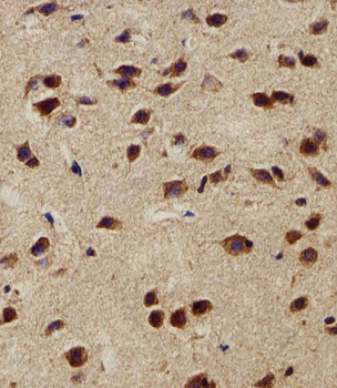
Immunohistochemical analysis of paraffin-embedded M. brain section using NEK2 Antibody (Center). Diluted at 1: 100 dilution. A peroxidase-conjugated goat anti-rabbit IgG at 1:400 dilution was used as the secondary antibody, followed by DAB staining.
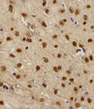
Immunohistochemical analysis of paraffin-embedded R. brain section using NEK2 Antibody (Center). Diluted at 1: 100 dilution. A peroxidase-conjugated goat anti-rabbit IgG at 1:400 dilution was used as the secondary antibody, followed by DAB staining.
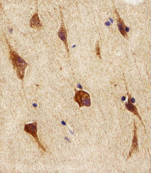
Immunohistochemical analysis of paraffin-embedded H. brain section using NEK2 Antibody (Center). Diluted at 1: 100 dilution. A peroxidase-conjugated goat anti-rabbit IgG at 1:400 dilution was used as the secondary antibody, followed by DAB staining.
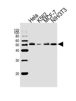
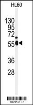
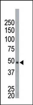
Western blot analysis of lysates from Hela, K562, MCF-7, mouse NIH/3T3 cell line (from left to right), using NEK2 Antibody (C410). Diluted at 1:1000 at each lane. A goat anti-rabbit IgG H&L (HRP) at 1:10000 dilution was used as the secondary antibody.
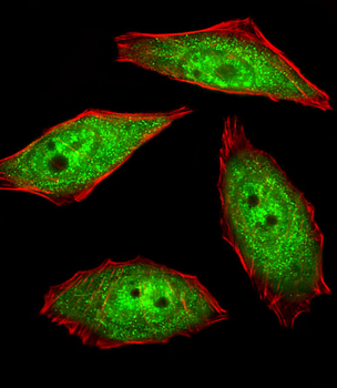
Fluorescent image of U251 cells stained with hNEK2-C410. Diluted at 1:25 dilution. An Alexa Fluor 488-conjugated goat anti-rabbit lgG at 1:400 dilution was used as the secondary antibody (green). Cytoplasmic actin was counterstained with Alexa Fluor 555 conjugated with Phalloidin (red).
Quick Database Links
Gene Symbol
This NEK2 antibody is generated from rabbits immunized with a KLH conjugated synthetic peptide between 396-426 amino acids from the Central region of human NEK2.
UniProt
RefSeq (Protein):NP_001191111.1, NP_002488.1, NP_001191112.1
UniProt Details
− No UniProt data available
NCBI Reference Sequences
−Associated Accession Numbers
Curated reference sequences for the gene transcript and protein product| Protein | NP_001191111.1, NP_002488.1, NP_001191112.1 |
|---|
Documents Download
Datasheet
Product Information
Request a Document
Protocol Information
WB
Western Blot (IB, immunoblot)
IHC-P
Immunohistochemistry Paraffin
IF
Immunofluorescence
NEK2 Antibody (Center) (orb1788313)

- 0.0
Based on 0 reviews
Participating in our Biorbyt product reviews program enables you to support fellow scientists by sharing your firsthand experience with our products.
Login to Submit a ReviewAvailable Sizes
Select a size below