You have no items in your shopping cart.
Featured
Description
Research Area
Alzheimer's Disease, Cell Biology, Host Immune Response, Host-Virus Interaction, Inflammation, Inflammatory Mediators, Innate Immunity, NF-kB Signalling Pathway, Neuroscience, Obesity, RELA (p65), Virology
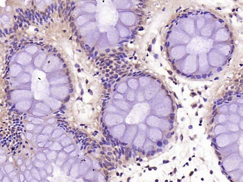
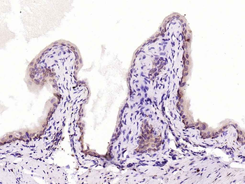
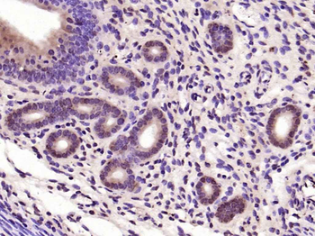
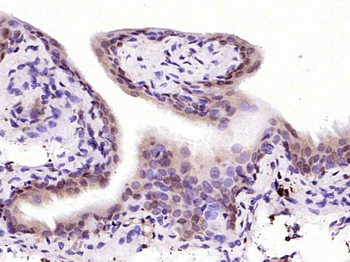
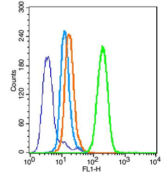
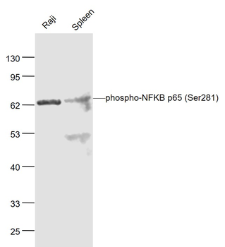
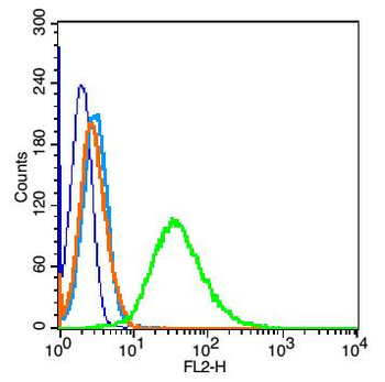
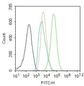
Images & Validation
−


Item 1 of 15
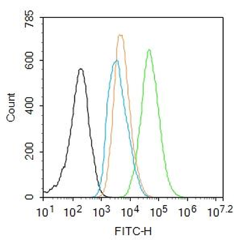
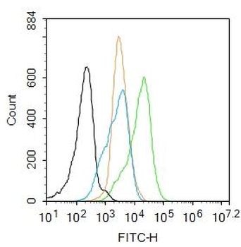
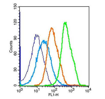
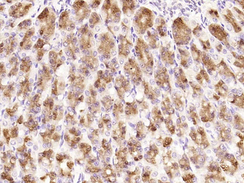
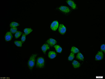
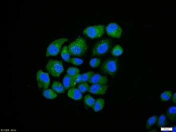
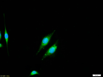
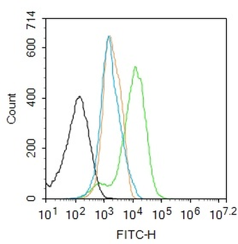
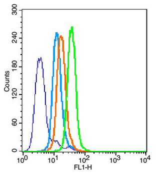
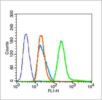
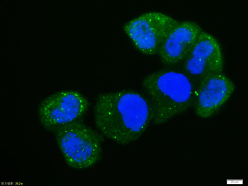
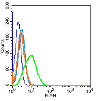
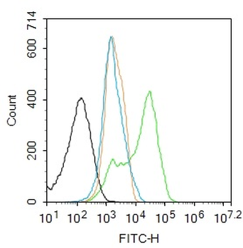
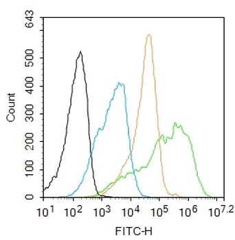
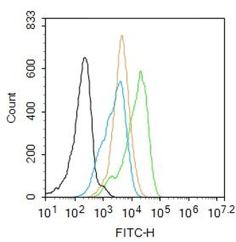
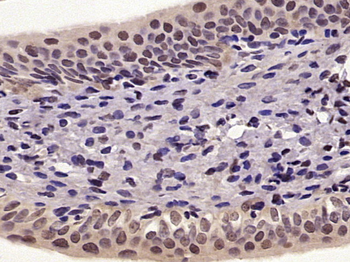
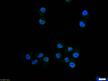
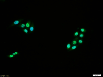
| Tested Applications | FC, ICC |
|---|---|
| Dilution Range | ICC/IF=1:50-200, Flow-Cyt=1μg/Test |
| Reactivity | Human, Mouse |
| Predicted Reactivity | Bovine, Canine, Equine, Gallus, Porcine, Rabbit, Rat, Sheep, Zebrafish |
Related Conjugates & Formulations
−Key Properties
−| Antibody Type | Primary Antibody |
|---|---|
| Host | Rabbit |
| Clonality | Polyclonal |
| Isotype | IgG |
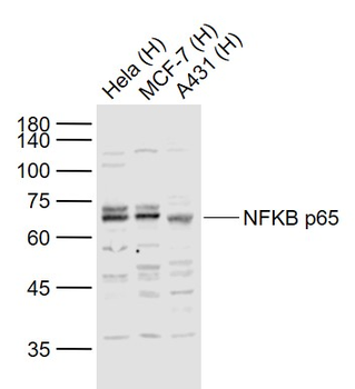
| Immunogen | KLH conjugated synthetic peptide derived from human NFKBp65 (51-100/551aa) |
| Target | RELA |
| Molecular Weight | 61 kDa |
| Purification | Affinity purified by Protein A |
| Conjugation | Unconjugated |
Storage & Handling
−| Storage | Maintain refrigerated at 2-8°C for up to 2 weeks. For long term storage store at -20°C in small aliquots to prevent freeze-thaw cycles. |
|---|---|
| Form/Appearance | Liquid |
| Buffer/Preservatives | 0.01M TBS (pH7.4) with 1% rAlbumin, 0.02% Proclin300 and 50% Glycerol. |
| Concentration | 1mg/ml |
| Expiration Date | 12 months from date of receipt. |
| Disclaimer | For research use only |
Alternative Names
−AIF3BL3; CMCU; NFKB3; p65; p65 NF-kappa B; p65 NFkB; NFkB; nos2; TF65_HUMAN; RELA; Nuclear factor NF-kappa-B p65 subunit; Nuclear factor of kappa light polypeptide gene enhancer in B-cells 3; TF65_MOUSE; RELA proto-oncogene, NF-kB subunit; v-rel avian reticuloendotheliosis viral oncogene homolog A
Similar Products
−- Item 1 of 9
NFKB p65 Rabbit Polyclonal Antibody [orb312399]
FC, ICC, IF, IHC-Fr, IHC-P, KO/KD Validated, WB
Bovine, Canine, Porcine
Human, Mouse, Rat
Rabbit
Polyclonal
Unconjugated
50 μl, 100 μl, 200 μl - Item 1 of 8
Phospho-NFKB p65 (Ser468) Rabbit Polyclonal Antibody [orb6503]
FC, ICC, IF, IHC-Fr, IHC-P, WB
Bovine, Canine, Equine, Porcine
Human, Mouse, Rat
Rabbit
Polyclonal
Unconjugated
50 μl, 100 μl, 200 μl - Item 1 of 6
Phospho-NFKB p65 (Ser536) Rabbit Polyclonal Antibody [orb6504]
WB
Human, Monkey, Rat
Mouse
Rabbit
Polyclonal
Unconjugated
50 μl, 100 μl, 200 μl - Item 1 of 7
Phospho-NFKB p65 (Ser281) Rabbit Polyclonal Antibody [orb185656]
FC, WB
Bovine, Canine, Equine, Porcine, Rat, Sheep
Human, Mouse
Rabbit
Polyclonal
Unconjugated
50 μl, 100 μl, 200 μl - Item 1 of 6
Phospho-NFKB p65 (Ser276) Rabbit Polyclonal Antibody [orb106211]
FC, ICC, IF, IHC-Fr, IHC-P
Bovine, Canine, Equine, Mouse, Porcine
Human, Rat
Rabbit
Polyclonal
Unconjugated
50 μl, 100 μl, 200 μl


Quality Guarantee
Explore bioreagents carefree to elevate your research. All our products are rigorously tested for performance. If a product does not perform as described on its datasheet, our scientific support team will provide expert troubleshooting, a prompt replacement, or a refund. For full details, please see our Terms & Conditions and Buying Guide. Contact us at support@biorbyt.com.
Quick Database Links
Gene Symbol
RELA
UniProt
UniProt Details
− No UniProt data available
Protocol Information
FC
Flow Cytometry
ICC
Immunocytochemistry
Filter by Applications
Filter by Species
Rituparna Ghosh 1, Biswadev Bishayi 2 Neutralization of TLR2 in combination with either TNF-α or IL-1β antibody reduces the severity of septic arthritis through STAT3/mTOR signalling in lymphocytes Cell Immunol ., (2024)
Applications
WB
Reactivity
Mouse
Sahin Sultana, Rajen Dey, Biswadev Bishayi Dual neutralization of TNFR-2 and MMP-2 regulates the severity of S. aureus induced septic arthritis correlating alteration in the level of interferon gamma and interleukin-10 in terms of TNFR2 blocking Immunologic Research, 66, 97 (2018)
Applications
WB
Reactivity
Mouse
Song, Yong et al. NF κB expression increases and CFTR and MUC1 expression decreases in the endometrium of infertile patients with hydrosalpinx: a comparative study Reprod Biol Endocrinol, 10, 86 (2012)
Available Sizes
Select a size below
Free Secondary Antibody (20 ul)0/0
Please add an antibody product to your cart first.