You have no items in your shopping cart.
Description
Research Area
Cancer Biology, Protein Biochemistry, Signal Transduction
Images & Validation
−

Item 1 of 3
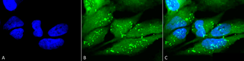
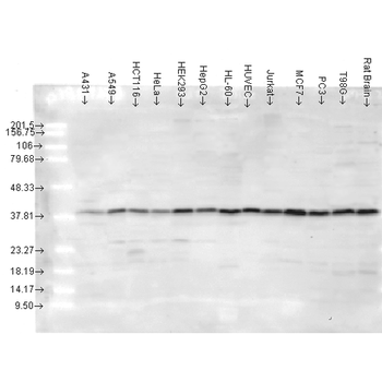
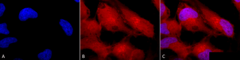
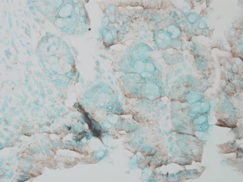
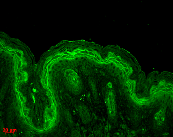
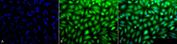
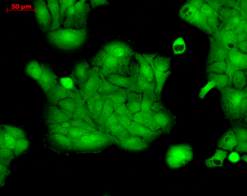
| Tested Applications | ICC, IF, IHC, IP, WB |
|---|---|
| Dilution Range | WB (1:1000), ICC/IF (1:100), IP (1:250) |
| Reactivity | Bovine, Canine, Gallus, Guinea pig, Hamster, Human, Monkey, Mouse, Porcine, Rabbit, Rat, Sheep |
| Application Notes |
Key Properties
−| Host | Rabbit |
|---|---|
| Clonality | Polyclonal |
| Immunogen | A 20 residue synthetic peptide based on the human p38 with the cysteine residue added and coupled to KLH |
| Target | p38 |
| Molecular Weight | 43kDa |
| Purification | Peptide Affinity Purified |
| Conjugation | Biotin |
Storage & Handling
−| Storage | Conjugated antibodies should be stored according to the product label |
|---|---|
| Buffer/Preservatives | 136.36mM Ethanolamine, 133.23 mM Chlorides, 9.55mM Phosphates, 9.55mM Sodium Bicarbonate. |
| Concentration | 1 mg/ml |
| Expiration Date | 12 months from date of receipt. |
| Disclaimer | For research use only |
Alternative Names
−CSAID Binding protein 1, CSBP1, CSBP2, EXIP, MAP kinase MXI2, MAPkinase p38alpha, MAPK14, p38 ALPHA, p38 MAP kinase, p38 mitogen activated protein kinase, RK, SAPK 2A, Stress activated protein kinase 2A
Similar Products
−- Item 1 of 6
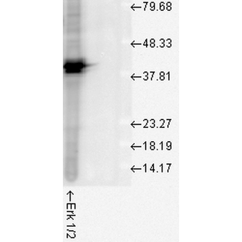
Erk1/2 Antibody (Biotin) [orb151496]
FC, ICC, IF, IHC, WB
Bovine, Drosophila, Frog, Gallus, Human, Mouse, Rat, Sheep
Rabbit
Polyclonal
Biotin
100 μl - Item 1 of 1
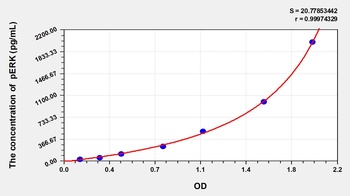
Rat Phospho Extracellular Signal Regulated Kinase (pERK) ELISA Kit [orb1088243]
Rat
31.25-2000 pg/mL
11.79 pg/mL
48 T, 96 T - Item 1 of 1
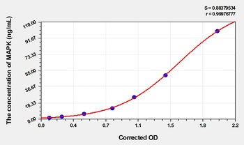
Human Mitogen Activated Protein Kinase (MAPK) ELISA Kit [orb1736622]
Human
1.57-100 ng/mL
0.53 ng/mL
48 T, 96 T - Item 1 of 1
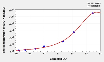
Rat P38 Mitogen Activated Protein Kinase (MAPK) ELISA Kit [orb1736653]
Rat
1.25-80 ng/mL
0.49 ng/mL
48 T, 96 T - Item 1 of 1
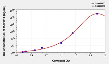
Dog Mitogen Activated Protein Kinase 14 (MAPK14) ELISA Kit [orb1817365]
Canine
0.32-20 ng/mL
0.12 ng/mL
48 T, 96 T
Quality Guarantee
Explore bioreagents carefree to elevate your research. All our products are rigorously tested for performance. If a product does not perform as described on its datasheet, our scientific support team will provide expert troubleshooting, a prompt replacement, or a refund. For full details, please see our Terms & Conditions and Buying Guide. Contact us at support@biorbyt.com.
Quick Database Links
UniProt Details
− No UniProt data available
NCBI Gene Details
− No NCBI Gene data available
NCBI Reference Sequences
−Associated Accession Numbers
Curated reference sequences for the gene transcript and protein product| Protein | NP_001306.1 |
|---|
Protocol Information
WB
Western Blot (IB, immunoblot)
IHC
Immunohistochemistry
IF
Immunofluorescence
ICC
Immunocytochemistry
IP
Immunoprecipitation
Available Sizes
Select a size below
Choose Conjugation or Carrier Free Version
Free Secondary Antibody (20 ul)0/0
Please add an antibody product to your cart first.