You have no items in your shopping cart.
Description
Research Area
Cell Biology
Images & Validation
−Item 1 of 7
| Tested Applications | FC, IF, IHC-P, WB |
|---|---|
| Dilution Range | IF - 1:100, WB - 1:1000, IHC-P - 1:100, FC - 1:10-50 |
| Reactivity | Human, Mouse, Rat |
Key Properties
−| Host | Rabbit |
|---|---|
| Clonality | Polyclonal |
| Isotype | Rabbit IgG |
| Immunogen | This PDIA6 antibody is generated from rabbits immunized with a KLH conjugated synthetic peptide between 144-172 amino acids from the Central region of human PDIA6. Antigen Region: 144-172 aa. |
| Target | PDIA6 |
| Molecular Weight | 48121 Da |
| Conjugation | Unconjugated |
Storage & Handling
−| Storage | Maintain refrigerated at 2-8°C for up to 2 weeks. For long term storage store at -20°C in small aliquots to prevent freeze-thaw cycles |
|---|---|
| Form/Appearance | Purified polyclonal antibody supplied in PBS with 0.09% (W/V) sodium azide. This antibody is prepared by Saturated Ammonium Sulfate (SAS) precipitation followed by dialysis against PBS. |
| Expiration Date | 12 months from date of receipt. |
| Disclaimer | For research use only |
Alternative Names
−Protein disulfide-isomerase A6, Endoplasmic reticulum protein 5, ER protein 5, ERp5, Protein disulfide isomerase P5, Thioredoxin domain-containing protein 7, PDIA6, ERP5, P5, TXNDC7
Quality Guarantee
Explore bioreagents carefree to elevate your research. All our products are rigorously tested for performance. If a product does not perform as described on its datasheet, our scientific support team will provide expert troubleshooting, a prompt replacement, or a refund. For full details, please see our Terms & Conditions and Buying Guide. Contact us at support@biorbyt.com.
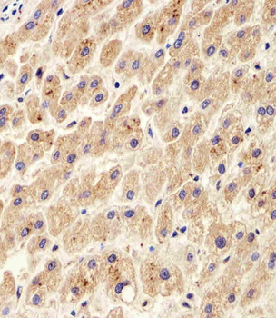
Immunohistochemical analysis of paraffin-embedded H. liver section using PDIA6 Antibody (Center K159). Diluted at 1:100 dilution. A peroxidase-conjugated goat anti-rabbit IgG at 1:400 dilution was used as the secondary antibody, followed by DAB staining.
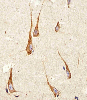
Immunohistochemical analysis of paraffin-embedded H. brain section using PDIA6 Antibody (Center K159). Diluted at 1:100 dilution. A peroxidase-conjugated goat anti-rabbit IgG at 1:400 dilution was used as the secondary antibody, followed by DAB staining.
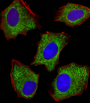
Fluorescent image of HepG2 cells stained with XAF1 PDIA6 Antibody (Center K159). Diluted at 1:100 dilution. An Alexa Fluor 488-conjugated goat anti-rabbit lgG at 1:400 dilution was used as the secondary antibody (green). DAPI was used to stain the cell nuclear (blue). Cytoplasmic actin was counterstained with Alexa Fluor 555 conjugated with Phalloidin (red).
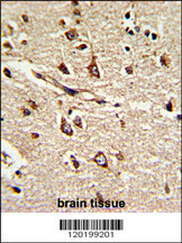
Formalin-fixed and paraffin-embedded human brain tissue reacted with PDIA6 Antibody (Center K159), which was peroxidase-conjugated to the secondary antibody, followed by DAB staining. This data demonstrates the use of this antibody for immunohistochemistry; clinical relevance has not been evaluated.
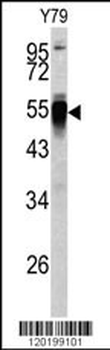
Western blot analysis of PDIA6 antibody (Center K159) in Y79 cell line lysates (35 ug/lane). PDIA6 (arrow) was detected using the purified Pab.
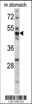
Western blot analysis of PDIA6 antibody (Center K159) in mouse stomach tissue lysates (35 ug/lane). PDIA6 (arrow) was detected using the purified Pab.
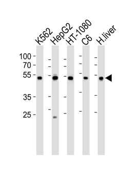
Western blot analysis of lysates from K562, HepG2, HT-1080, rat C6 cell line and human liver tissue lysate (from left to right), using PDIA6 Antibody (Center K159). Diluted at 1:1000 at each lane. A goat anti-rabbit IgG H&L (HRP) at 1:10000 dilution was used as the secondary antibody. Lysates at 35 ug per lane.
Quick Database Links
UniProt Details
− No UniProt data available
NCBI Reference Sequences
−Associated Accession Numbers
Curated reference sequences for the gene transcript and protein product| Protein | NP_005733.1 |
|---|
Documents Download
Datasheet
Product Information
Request a Document
Protocol Information
WB
Western Blot (IB, immunoblot)
IHC-P
Immunohistochemistry Paraffin
FC
Flow Cytometry
IF
Immunofluorescence
PDIA6 Antibody (Center K159) (orb1929905)

- 0.0
Based on 0 reviews
Participating in our Biorbyt product reviews program enables you to support fellow scientists by sharing your firsthand experience with our products.
Login to Submit a ReviewAvailable Sizes
Select a size below
Choose Conjugation or Carrier Free Version
Free Secondary Antibody (20 ul)0/0
Please add an antibody product to your cart first.